


New Super-Slow Substrates as novel Sirtuin-Inhibitors
Friedrich, F., Kalbas, D., Einsle, O., Jung, M., Schutkowski, M.To be published.
Experimental Data Snapshot
Starting Model: experimental
View more details
Entity ID: 1 | |||||
|---|---|---|---|---|---|
| Molecule | Chains | Sequence Length | Organism | Details | Image |
| NAD-dependent protein deacetylase sirtuin-2 | 304 | Homo sapiens | Mutation(s): 0 Gene Names: SIRT2, SIR2L, SIR2L2 EC: 3.5.1 (PDB Primary Data), 2.3.1 (UniProt), 2.3.1.286 (UniProt) | 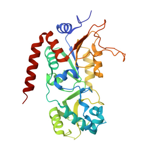 | |
UniProt & NIH Common Fund Data Resources | |||||
GTEx: ENSG00000068903 | |||||
Entity Groups | |||||
| Sequence Clusters | 30% Identity50% Identity70% Identity90% Identity95% Identity100% Identity | ||||
| UniProt Group | Q8IXJ6 | ||||
Sequence AnnotationsExpand | |||||
Reference Sequence | |||||
Entity ID: 2 | |||||
|---|---|---|---|---|---|
| Molecule | Chains | Sequence Length | Organism | Details | Image |
| Peptide-based super-slow substrate TNFn-3 | 10 | Homo sapiens | Mutation(s): 0 | 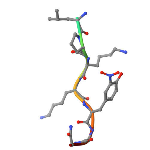 | |
Entity Groups | |||||
| Sequence Clusters | 30% Identity50% Identity70% Identity90% Identity95% Identity100% Identity | ||||
Sequence AnnotationsExpand | |||||
Reference Sequence | |||||
| Ligands 4 Unique | |||||
|---|---|---|---|---|---|
| ID | Chains | Name / Formula / InChI Key | 2D Diagram | 3D Interactions | |
| WWE (Subject of Investigation/LOI) Download:Ideal Coordinates CCD File | H [auth B] | 3-dodecylsulfanyl-3-methyl-butanoic acid C17 H34 O2 S FVOHTIJLQOWWCZ-UHFFFAOYSA-N |  | ||
| PEG Download:Ideal Coordinates CCD File | D [auth A] | DI(HYDROXYETHYL)ETHER C4 H10 O3 MTHSVFCYNBDYFN-UHFFFAOYSA-N |  | ||
| ZN Download:Ideal Coordinates CCD File | C [auth A] | ZINC ION Zn PTFCDOFLOPIGGS-UHFFFAOYSA-N |  | ||
| EDO Download:Ideal Coordinates CCD File | E [auth A], F [auth A], G [auth A] | 1,2-ETHANEDIOL C2 H6 O2 LYCAIKOWRPUZTN-UHFFFAOYSA-N |  | ||
| Modified Residues 1 Unique | |||||
|---|---|---|---|---|---|
| ID | Chains | Type | Formula | 2D Diagram | Parent |
| NIY Query on NIY | B | L-PEPTIDE LINKING | C9 H10 N2 O5 |  | TYR |
| Length ( Å ) | Angle ( ˚ ) |
|---|---|
| a = 36.144 | α = 90 |
| b = 74.318 | β = 94.28 |
| c = 56.052 | γ = 90 |
| Software Name | Purpose |
|---|---|
| PHENIX | refinement |
| Aimless | data scaling |
| autoPROC | data reduction |
| PHASER | phasing |
| Funding Organization | Location | Grant Number |
|---|---|---|
| German Research Foundation (DFG) | Germany | SFB 992 |
| German Research Foundation (DFG) | Germany | 295/18-1 |


