8QQE

Crystal structure of the complex between DMC1 and the PhePP domain of BRCA2


Experimental Data Snapshot

  • Method: X-RAY DIFFRACTION
  • Resolution: 3.46 Å
  • R-Value Free: 0.261 
  • R-Value Work: 0.248 
  • R-Value Observed: 0.249 

wwPDB Validation   3D Report Full Report


This is version 1.0 of the entry. See complete history


Literature

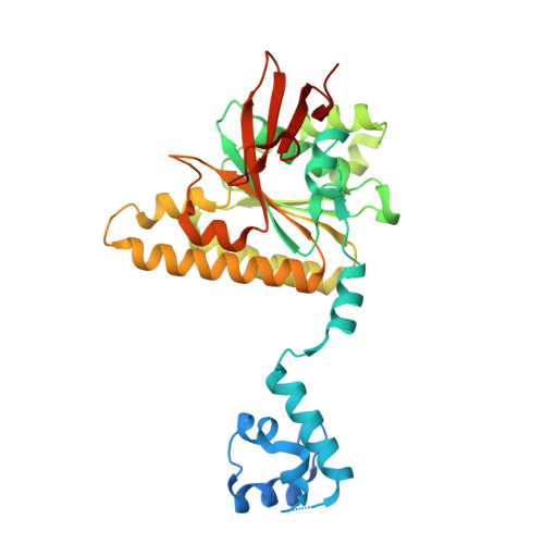
DMC1 and RAD51 bind FxxA and FxPP motifs of BRCA2 via two separate interfaces.

Miron, S.Legrand, P.Dupaigne, P.van Rossum-Fikkert, S.E.Ristic, D.Majeed, A.Kanaar, R.Zinn-Justin, S.Zelensky, A.N.

(2024) Nucleic Acids Res 

  • DOI: https://doi.org/10.1093/nar/gkae452
  • Primary Citation of Related Structures:  
    8QQE

  • PubMed Abstract: 

    In vertebrates, the BRCA2 protein is essential for meiotic and somatic homologous recombination due to its interaction with the RAD51 and DMC1 recombinases through FxxA and FxPP motifs (here named A- and P-motifs, respectively). The A-motifs present in the eight BRC repeats of BRCA2 compete with the A-motif of RAD51, which is responsible for its self-oligomerization. BRCs thus disrupt RAD51 nucleoprotein filaments in vitro. The role of the P-motifs is less studied. We recently found that deletion of Brca2 exons 12-14 encoding one of them (the prototypical 'PhePP' motif), disrupts DMC1 but not RAD51 function in mouse meiosis. Here we provide a mechanistic explanation for this phenotype by solving the crystal structure of the complex between a BRCA2 fragment containing the PhePP motif and DMC1. Our structure reveals that, despite sharing a conserved phenylalanine, the A- and P-motifs bind to distinct sites on the ATPase domain of the recombinases. The P-motif interacts with a site that is accessible in DMC1 octamers and nucleoprotein filaments. Moreover, we show that this interaction also involves the adjacent protomer and thus increases the stability of the DMC1 nucleoprotein filaments. We extend our analysis to other P-motifs from RAD51AP1 and FIGNL1.


  • Organizational Affiliation

    Institute for Integrative Biology of the Cell (I2BC), Université Paris-Saclay, CEA, CNRS, Gif-sur-Yvette, France.


Macromolecules
Find similar proteins by:  (by identity cutoff)  |  3D Structure
Entity ID: 1
MoleculeChains Sequence LengthOrganismDetailsImage
Meiotic recombination protein DMC1/LIM15 homolog
A, B
340Homo sapiensMutation(s): 0 
Gene Names: DMC1
UniProt & NIH Common Fund Data Resources
Find proteins for Q14565 (Homo sapiens)
Explore Q14565 
Go to UniProtKB:  Q14565
PHAROS:  Q14565
GTEx:  ENSG00000100206 
Entity Groups  
Sequence Clusters30% Identity50% Identity70% Identity90% Identity95% Identity100% Identity
UniProt GroupQ14565
Sequence Annotations
Expand
  • Reference Sequence

Find similar proteins by:  Sequence   |   3D Structure  

Entity ID: 2
MoleculeChains Sequence LengthOrganismDetailsImage
Breast cancer type 2 susceptibility protein
C, D
20Homo sapiensMutation(s): 0 
Gene Names: BRCA2
UniProt & NIH Common Fund Data Resources
Find proteins for P51587 (Homo sapiens)
Explore P51587 
Go to UniProtKB:  P51587
PHAROS:  P51587
GTEx:  ENSG00000139618 
Entity Groups  
Sequence Clusters30% Identity50% Identity70% Identity90% Identity95% Identity100% Identity
UniProt GroupP51587
Sequence Annotations
Expand
  • Reference Sequence
Experimental Data & Validation

Experimental Data

  • Method: X-RAY DIFFRACTION
  • Resolution: 3.46 Å
  • R-Value Free: 0.261 
  • R-Value Work: 0.248 
  • R-Value Observed: 0.249 
  • Space Group: I 4 3 2
Unit Cell:
Length ( Å )Angle ( ˚ )
a = 234.383α = 90
b = 234.383β = 90
c = 234.383γ = 90
Software Package:
Software NamePurpose
BUSTERrefinement
MxCuBEdata collection
XDSdata reduction
MOLREPphasing
STARANISOdata scaling

Structure Validation

View Full Validation Report


Entry History & Funding Information

Deposition Data


Funding OrganizationLocationGrant Number
Agence Nationale de la Recherche (ANR)FranceANR-10-INSB-05-01
iNEXT-DiscoveryEuropean Union871037

Revision History  (Full details and data files)

  • Version 1.0: 2024-06-19
    Type: Initial release