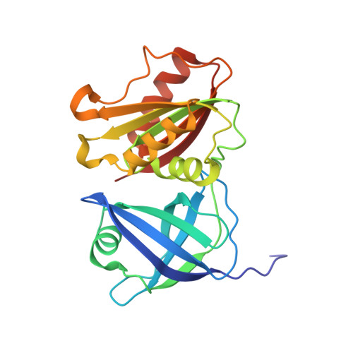
X-ray structure and enzymatic study of a bacterial NADPH oxidase highlight the activation mechanism of eukaryotic NOX.
Petit-Hartlein, I., Vermot, A., Thepaut, M., Humm, A.S., Dupeux, F., Dupuy, J., Chaptal, V., Marquez, J.A., Smith, S.M.E., Fieschi, F.(2024) Elife 13
- PubMed: 38640072 Search on PubMedSearch on PubMed Central
- DOI: https://doi.org/10.7554/eLife.93759
- Primary Citation Related Structures:
8QQ1, 8QQ5, 8QQ7 - PubMed Abstract:
NADPH oxidases (NOX) are transmembrane proteins, widely spread in eukaryotes and prokaryotes, that produce reactive oxygen species (ROS). Eukaryotes use the ROS products for innate immune defense and signaling in critical (patho)physiological processes. Despite the recent structures of human NOX isoforms, the activation of electron transfer remains incompletely understood. SpNOX, a homolog from Streptococcus pneumoniae , can serves as a robust model for exploring electron transfers in the NOX family thanks to its constitutive activity. Crystal structures of SpNOX full-length and dehydrogenase (DH) domain constructs are revealed here. The isolated DH domain acts as a flavin reductase, and both constructs use either NADPH or NADH as substrate. Our findings suggest that hydride transfer from NAD(P)H to FAD is the rate-limiting step in electron transfer. We identify significance of F397 in nicotinamide access to flavin isoalloxazine and confirm flavin binding contributions from both DH and Transmembrane (TM) domains. Comparison with related enzymes suggests that distal access to heme may influence the final electron acceptor, while the relative position of DH and TM does not necessarily correlate with activity, contrary to previous suggestions. It rather suggests requirement of an internal rearrangement, within the DH domain, to switch from a resting to an active state. Thus, SpNOX appears to be a good model of active NOX2, which allows us to propose an explanation for NOX2's requirement for activation.
- Univ. Grenoble Alpes, CNRS, CEA, Institut de Biologie Structurale, Grenoble, France.
Organizational Affiliation: