Mutational and structural studies of ( beta alpha ) 8 -barrel fold methylene-tetrahydropterin reductases utilizing a common catalytic mechanism.
Gehl, M., Demmer, U., Ermler, U., Shima, S.(2024) Protein Sci 33: e5018-e5018
- PubMed: 38747406 Search on PubMedSearch on PubMed Central
- DOI: https://doi.org/10.1002/pro.5018
- Primary Citation Related Structures:
8QPJ, 8QPL, 8QPM, 8QQ8 - PubMed Abstract:
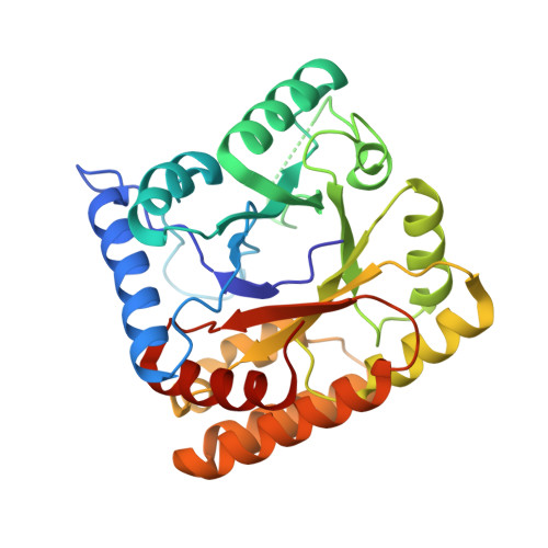
Methylene-tetrahydropterin reductases catalyze the reduction of a methylene to a methyl group bound to a reduced pterin as C 1 carrier in various one-carbon (C 1 ) metabolisms. F 420 -dependent methylene-tetrahydromethanopterin (methylene-H 4 MPT) reductase (Mer) and the flavin-independent methylene-tetrahydrofolate (methylene-H 4 F) reductase (Mfr) use a ternary complex mechanism for the direct transfer of a hydride from F 420 H 2 and NAD(P)H to the respective methylene group, whereas FAD-dependent methylene-H 4 F reductase (MTHFR) uses FAD as prosthetic group and a ping-pong mechanism to catalyze the reduction of methylene-H 4 F. A ternary complex structure and a thereof derived catalytic mechanism of MTHFR is available, while no ternary complex structures of Mfr or Mer are reported. Here, Mer from Methanocaldococcus jannaschii (jMer) was heterologously produced and the crystal structures of the enzyme with and without F 420 were determined. A ternary complex of jMer was modeled on the basis of the jMer-F 420 structure and the ternary complex structure of MTHFR by superimposing the polypeptide after fixing hydride-transferring atoms of the flavins on each other, and by the subsequent transfer of the methyl-tetrahydropterin from MTHFR to jMer. Mutational analysis of four functional amino acids, which are similarly positioned in the three reductase structures, indicated despite the insignificant sequence identity, a common catalytic mechanism with a 5-iminium cation of methylene-tetrahydropterin as intermediate protonated by a shared glutamate. According to structural, mutational and phylogenetic analysis, the evolution of the three reductases most likely proceeds via a convergent development although a divergent scenario requiring drastic structural changes of the common ancestor cannot be completely ruled out.
- Max Planck Institute for Terrestrial Microbiology, Marburg, Germany.
Organizational Affiliation: