Three-dimensional solution structure, dynamics and binding of thioredoxin m from Pisum sativum.
Neira, J.L., Palomino-Schatzlein, M., Rejas, V., Traverso, J.A., Rico, M., Lopez-Gorge, J., Chueca, A., Camara-Artigas, A.(2024) Int J Biol Macromol 262: 129781-129781
- PubMed: 38296131 Search on PubMed
- DOI: https://doi.org/10.1016/j.ijbiomac.2024.129781
- Primary Citation Related Structures:
8QPD - PubMed Abstract:
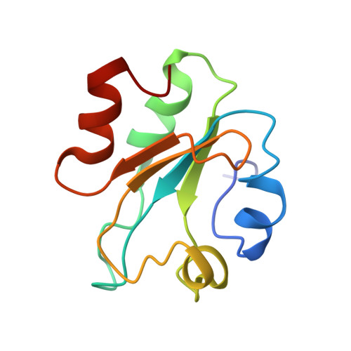
Thioredoxins (TRXs) are ubiquitous small, globular proteins involved in cell redox processes. In this work, we report the solution structure of TRX m from Pisum sativum (pea), which has been determined on the basis of 1444 nuclear Overhauser effect- (NOE-) derived distance constraints. The average pairwise root-mean-square deviation (RMSD) for the 20 best structures for the backbone residues (Val7-Glu102) was 1.42 ± 0.15 Å, and 1.97 ± 0.15 Å when all heavy atoms were considered. The structure corresponds to the typical fold of TRXs, with a central five-stranded β-sheet flanked by four α-helices. Some residues had an important exchange dynamic contribution: those around the active site; at the C terminus of β-strand 3; and in the loop preceding α-helix 4. Smaller NOE values were observed at the N and C-terminal residues forming the elements of the secondary structure or, alternatively, in the residues belonging to the loops between those elements. A peptide derived from pea fructose-1,6-biphosphatase (FBPase), comprising the preceding region to the regulatory sequence of FBPase (residues Glu152 to Gln179), was bound to TRX m with an affinity in the low micromolar range, as measured by fluorescence and NMR titration experiments. Upon peptide addition, the intensities of the cross-peaks of all the residues of TRX m were affected, as shown by NMR. The value of the dissociation constant of the peptide from TRX m was larger than that of the intact FBPase, indicating that there are additional factors in other regions of the polypeptide chain of the latter protein affecting the binding to thioredoxin.
- IDIBE, Universidad Miguel Hernández, 03202 Elche, Alicante, Spain; Instituto de Biocomputación y Física de Sistemas Complejos (BIFI), Universidad de Zaragoza, 50018 Zaragoza, Spain. Electronic address: jlneira@umh.es.
Organizational Affiliation: