8QJG | pdb_00008qjg

Crystal structure of cytochrome domain 2 from PgcA


Experimental Data Snapshot

  • Method: X-RAY DIFFRACTION
  • Resolution: 1.80 Å
  • R-Value Free: 
    0.227 (Depositor), 0.232 (DCC) 
  • R-Value Work: 
    0.175 (Depositor), 0.183 (DCC) 
  • R-Value Observed: 
    0.177 (Depositor) 

wwPDB Validation 3D Report Full Report

Validation slider image for 8QJG

Ligand Structure Quality Assessment 


This is version 1.0 of the entry. See complete history

Literature

Tethered heme domains in a triheme cytochrome allow for increased electron transport distances.

Nash, B.W.Fernandes, T.M.Burton, J.A.J.Morgado, L.van Wonderen, J.H.Svistunenko, D.A.Edwards, M.J.Salgueiro, C.A.Butt, J.N.Clarke, T.A.

(2024) Protein Sci 33: e5200-e5200

  • DOI: https://doi.org/10.1002/pro.5200
  • Primary Citation Related Structures: 
    8QJ6, 8QJG, 8QK0

  • PubMed Abstract: 

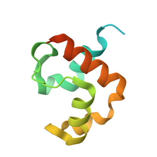
    Decades of research describe myriad redox enzymes that contain cofactors arranged in tightly packed chains facilitating rapid and controlled intra-protein electron transfer. Many such enzymes participate in extracellular electron transfer (EET), a process which allows microorganisms to conserve energy in anoxic environments by exploiting mineral oxides and other extracellular substrates as terminal electron acceptors. In this work, we describe the properties of the triheme cytochrome PgcA from Geobacter sulfurreducens. PgcA has been shown to play an important role in EET but is unusual in containing three CXXCH heme binding motifs that are separated by repeated (PT) x motifs, suggested to enhance binding to mineral surfaces. Using a combination of structural, electrochemical, and biophysical techniques, we experimentally demonstrate that PgcA adopts numerous conformations stretching as far as 180 Å between the ends of domains I and III, without a tightly packed cofactor chain. Furthermore, we demonstrate a distinct role for its domain III as a mineral reductase that is recharged by domains I and II. These findings show PgcA to be the first of a new class of electron transfer proteins, with redox centers separated by some nanometers but tethered together by flexible linkers, facilitating electron transfer through a tethered diffusion mechanism rather than a fixed, closely packed electron transfer chain.


  • Organizational Affiliation
    • Centre for Molecular and Structural Biochemistry, School of Biological Sciences and School of Chemistry, University of East Anglia, Norwich, UK.

Macromolecule Content 

  • Total Structure Weight: 64.89 kDa 
  • Atom Count: 3,555 
  • Modeled Residue Count: 391 
  • Deposited Residue Count: 588 
  • Unique protein chains: 1

Macromolecules

Find similar proteins by:|  3D Structure
Entity ID: 1
MoleculeChains  Sequence LengthOrganismDetailsImage
Lipoprotein cytochrome c
A, B, C, D, E
A, B, C, D, E, F
98Geobacter sulfurreducens PCAMutation(s): 0 
Gene Names: pgcA
UniProt
Find proteins for Q74CB3 (Geobacter sulfurreducens (strain ATCC 51573 / DSM 12127 / PCA))
Explore Q74CB3 
Go to UniProtKB:  Q74CB3
Entity Groups
Sequence Clusters30% Identity50% Identity70% Identity90% Identity95% Identity100% Identity
UniProt GroupQ74CB3
Sequence Annotations
Expand
Reference Sequence

Small Molecules

Ligands 3 Unique
IDChains Name / Formula / InChI Key2D Diagram3D Interactions
HEC
(Subject of Investigation/LOI)

Query on HEC


Download:Ideal Coordinates CCD File
I [auth A]
K [auth B]
O [auth C]
S [auth D]
V [auth E]
I [auth A],
K [auth B],
O [auth C],
S [auth D],
V [auth E],
Z [auth F]
HEME C
C34 H34 Fe N4 O4
HXQIYSLZKNYNMH-LJNAALQVSA-N
SO4

Query on SO4


Download:Ideal Coordinates CCD File
G [auth A]
H [auth A]
J [auth B]
L [auth C]
M [auth C]
G [auth A],
H [auth A],
J [auth B],
L [auth C],
M [auth C],
N [auth C],
P [auth D],
Q [auth D],
R [auth D],
T [auth E],
U [auth E],
W [auth F],
X [auth F]
SULFATE ION
O4 S
QAOWNCQODCNURD-UHFFFAOYSA-L
EDO

Query on EDO


Download:Ideal Coordinates CCD File
Y [auth F]1,2-ETHANEDIOL
C2 H6 O2
LYCAIKOWRPUZTN-UHFFFAOYSA-N

Experimental Data & Validation

Experimental Data

  • Method: X-RAY DIFFRACTION
  • Resolution: 1.80 Å
  • R-Value Free:  0.227 (Depositor), 0.232 (DCC) 
  • R-Value Work:  0.175 (Depositor), 0.183 (DCC) 
  • R-Value Observed: 0.177 (Depositor) 
Space Group: C 1 2 1
Unit Cell:
Length ( Å )Angle ( ˚ )
a = 95.104α = 90
b = 74.101β = 103.92
c = 56.019γ = 90
Software Package:
Software NamePurpose
PHENIXrefinement
Aimlessdata scaling
xia2data reduction
MOLREPphasing

Structure Validation

View Full Validation Report


Ligand Structure Quality Assessment 


Entry History 

& Funding Information

Deposition Data


Funding OrganizationLocationGrant Number
Biotechnology and Biological Sciences Research Council (BBSRC)United KingdomBB/T008717/1

Revision History  (Full details and data files)

  • Version 1.0: 2024-11-06
    Type: Initial release