Biological and structural investigation of tetrahydro-beta-carboline-based selective HDAC6 inhibitors with improved stability.
Scheuerer, S., Motlova, L., Schaker-Hubner, L., Sellmer, A., Feller, F., Ertl, F.J., Koch, P., Hansen, F.K., Barinka, C., Mahboobi, S.(2024) Eur J Med Chem 276: 116676-116676
- PubMed: 39067437 Search on PubMed
- DOI: https://doi.org/10.1016/j.ejmech.2024.116676
- Primary Citation Related Structures:
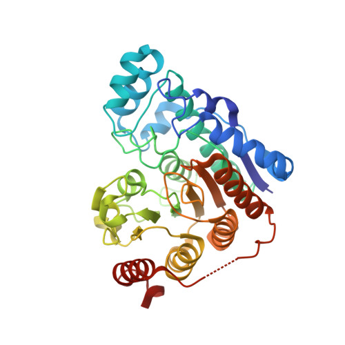
8QH9 - PubMed Abstract:
Our previously reported HDAC6 inhibitor (HDAC6i) Marbostat-100 (4) has provided many arguments for further clinical evaluation. By the substitution of the acidic hydrogen of 4 for different carbon residues, we were able to generate an all-carbon stereocenter, which significantly improves the hydrolytic stability of the inhibitor. Further asymmetric synthesis has shown that the (S)-configured inhibitors preferentially bind to HDAC6. This led to the highly selective and potent methyl-substituted derivative S-29b, which elicited a long-lasting tubulin hyperacetylation in MV4-11 cells. Finally, a crystal structure of the HDAC6/S-29b complex provided mechanistic explanation for the high potency and stereoselectivity of synthesized compound series.
- Institute of Pharmacy, Department of Pharmaceutical/Medicinal Chemistry I, University of Regensburg, 93040, Regensburg, Germany.
Organizational Affiliation: