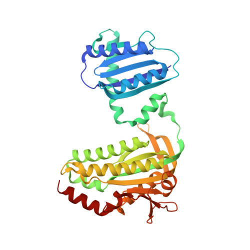
Light-induced Trp in /Met out Switching During BLUF Domain Activation in ATP-bound Photoactivatable Adenylate Cyclase OaPAC.
Chretien, A., Nagel, M.F., Botha, S., de Wijn, R., Brings, L., Dorner, K., Han, H., Koliyadu, J.C.P., Letrun, R., Round, A., Sato, T., Schmidt, C., Secareanu, R.C., von Stetten, D., Vakili, M., Wrona, A., Bean, R., Mancuso, A., Schulz, J., Pearson, A.R., Kottke, T., Lorenzen, K., Schubert, R.(2024) J Mol Biology 436: 168439-168439
- PubMed: 38185322 Search on PubMed
- DOI: https://doi.org/10.1016/j.jmb.2024.168439
- Primary Citation Related Structures:
8QFE, 8QFF, 8QFG, 8QFH, 8QFI, 8QFJ - PubMed Abstract:
The understanding of signal transduction mechanisms in photoreceptor proteins is essential for elucidating how living organisms respond to light as environmental stimuli. In this study, we investigated the ATP binding, photoactivation and signal transduction process in the photoactivatable adenylate cyclase from Oscillatoria acuminata (OaPAC) upon blue light excitation. Structural models with ATP bound in the active site of native OaPAC at cryogenic as well as room temperature are presented. ATP is found in one conformation at cryogenic- and in two conformations at ambient-temperature, and is bound in an energetically unfavorable conformation for the conversion to cAMP. However, FTIR spectroscopic experiments confirm that this conformation is the native binding mode in dark state OaPAC and that transition to a productive conformation for ATP turnover only occurs after light activation. A combination of time-resolved crystallography experiments at synchrotron and X-ray Free Electron Lasers sheds light on the early events around the Flavin Adenine Dinucleotide (FAD) chromophore in the light-sensitive BLUF domain of OaPAC. Early changes involve the highly conserved amino acids Tyr6, Gln48 and Met92. Crucially, the Gln48 side chain performs a 180° rotation during activation, leading to the stabilization of the FAD chromophore. Cryo-trapping experiments allowed us to investigate a late light-activated state of the reaction and revealed significant conformational changes in the BLUF domain around the FAD chromophore. In particular, a Trp in /Met out transition upon illumination is observed for the first time in the BLUF domain and its role in signal transmission via α-helix 3 and 4 in the linker region between sensor and effector domain is discussed.
- European XFEL GmbH, Schenefeld, Germany; Department of Chemistry, Universität Hamburg, Hamburg, Germany.
Organizational Affiliation: