8QAE

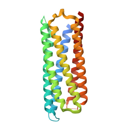
X-ray crystal structure of a de novo designed single-chain antiparallel 6-helix alpha-helical barrel, sc-apCC-6-SLLA


Experimental Data Snapshot

  • Method: X-RAY DIFFRACTION
  • Resolution: 1.90 Å
  • R-Value Free: 0.228 
  • R-Value Work: 0.206 
  • R-Value Observed: 0.207 

Starting Model: in silico
View more details

wwPDB Validation   3D Report Full Report


Ligand Structure Quality Assessment 


This is version 1.1 of the entry. See complete history


Literature

Rationally seeded computational protein design of ɑ-helical barrels.

Albanese, K.I.Petrenas, R.Pirro, F.Naudin, E.A.Borucu, U.Dawson, W.M.Scott, D.A.Leggett, G.J.Weiner, O.D.Oliver, T.A.A.Woolfson, D.N.

(2024) Nat Chem Biol 20: 991-999

  • DOI: https://doi.org/10.1038/s41589-024-01642-0
  • Primary Citation of Related Structures:  
    8QAA, 8QAB, 8QAC, 8QAD, 8QAE, 8QAF, 8QAG, 8QAH, 8QAI, 8QKD

  • PubMed Abstract: 

    Computational protein design is advancing rapidly. Here we describe efficient routes starting from validated parallel and antiparallel peptide assemblies to design two families of α-helical barrel proteins with central channels that bind small molecules. Computational designs are seeded by the sequences and structures of defined de novo oligomeric barrel-forming peptides, and adjacent helices are connected by loop building. For targets with antiparallel helices, short loops are sufficient. However, targets with parallel helices require longer connectors; namely, an outer layer of helix-turn-helix-turn-helix motifs that are packed onto the barrels. Throughout these computational pipelines, residues that define open states of the barrels are maintained. This minimizes sequence sampling, accelerating the design process. For each of six targets, just two to six synthetic genes are made for expression in Escherichia coli. On average, 70% of these genes express to give soluble monomeric proteins that are fully characterized, including high-resolution structures for most targets that match the design models with high accuracy.


  • Organizational Affiliation

    School of Chemistry, University of Bristol, Bristol, UK.


Macromolecules
Find similar proteins by:  (by identity cutoff)  |  3D Structure
Entity ID: 1
MoleculeChains Sequence LengthOrganismDetailsImage
sc-apCC-6-SLLAA,
B [auth C]
234synthetic constructMutation(s): 0 
Entity Groups  
Sequence Clusters30% Identity50% Identity70% Identity90% Identity95% Identity100% Identity
Sequence Annotations
Expand
  • Reference Sequence
Small Molecules
Ligands 2 Unique
IDChains Name / Formula / InChI Key2D Diagram3D Interactions
P5R
Query on P5R

Download Ideal Coordinates CCD File 
C [auth A]2-[2-[2-[2-[2-[2-[2-[2-[2-[2-[2-[2-[2-[2-[2-[2-(2-hydroxyethyloxy)ethoxy]ethoxy]ethoxy]ethoxy]ethoxy]ethoxy]ethoxy]ethoxy]ethoxy]ethoxy]ethoxy]ethoxy]ethoxy]ethoxy]ethoxy]ethanol
C34 H70 O18
NIELXDCPHZJHGM-UHFFFAOYSA-N
1PE
Query on 1PE

Download Ideal Coordinates CCD File 
D [auth C]PENTAETHYLENE GLYCOL
C10 H22 O6
JLFNLZLINWHATN-UHFFFAOYSA-N
Experimental Data & Validation

Experimental Data

  • Method: X-RAY DIFFRACTION
  • Resolution: 1.90 Å
  • R-Value Free: 0.228 
  • R-Value Work: 0.206 
  • R-Value Observed: 0.207 
  • Space Group: C 1 2 1
Unit Cell:
Length ( Å )Angle ( ˚ )
a = 126.823α = 90
b = 42.176β = 123.509
c = 71.522γ = 90
Software Package:
Software NamePurpose
PHENIXrefinement
DIALSdata reduction
DIALSdata scaling
PHASERphasing

Structure Validation

View Full Validation Report


Ligand Structure Quality Assessment 


Entry History & Funding Information

Deposition Data


Funding OrganizationLocationGrant Number
Max Planck Bristol Centre for Minimal Biology - University of BristolUnited Kingdom--

Revision History  (Full details and data files)

  • Version 1.0: 2024-07-03
    Type: Initial release
  • Version 1.1: 2024-08-07
    Changes: Database references