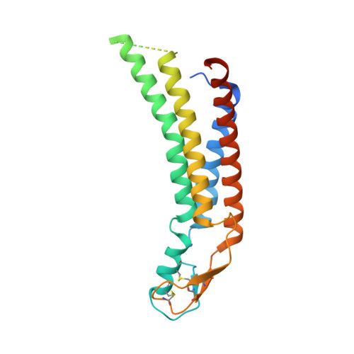
8QA1

Cryo-EM structure of Cx26 solubilised in LMNG - Hemichannel classification NFlex conformation


Experimental Data Snapshot

  • Method: ELECTRON MICROSCOPY
  • Resolution: 2.30 Å
  • Aggregation State: PARTICLE 
  • Reconstruction Method: SINGLE PARTICLE 

Starting Model: experimental
View more details

wwPDB Validation   3D Report Full Report


This is version 1.2 of the entry. See complete history


Literature

Structures of wild-type and a constitutively closed mutant of connexin26 shed light on channel regulation by CO 2.

Brotherton, D.H.Nijjar, S.Savva, C.G.Dale, N.Cameron, A.D.

(2024) Elife 13

  • DOI: https://doi.org/10.7554/eLife.93686
  • Primary Citation of Related Structures:  
    8Q9Z, 8QA0, 8QA1, 8QA2, 8QA3

  • PubMed Abstract: 

    Connexins allow intercellular communication by forming gap junction channels (GJCs) between juxtaposed cells. Connexin26 (Cx26) can be regulated directly by CO 2 . This is proposed to be mediated through carbamylation of K125. We show that mutating K125 to glutamate, mimicking the negative charge of carbamylation, causes Cx26 GJCs to be constitutively closed. Through cryo-EM we observe that the K125E mutation pushes a conformational equilibrium towards the channel having a constricted pore entrance, similar to effects seen on raising the partial pressure of CO 2 . In previous structures of connexins, the cytoplasmic loop, important in regulation and where K125 is located, is disordered. Through further cryo-EM studies we trap distinct states of Cx26 and observe density for the cytoplasmic loop. The interplay between the position of this loop, the conformations of the transmembrane helices and the position of the N-terminal helix, which controls the aperture to the pore, provides a mechanism for regulation.


  • Organizational Affiliation

    School of Life Sciences, University of Warwick, Coventry, United Kingdom.


Macromolecules
Find similar proteins by:  (by identity cutoff)  |  3D Structure
Entity ID: 1
MoleculeChains Sequence LengthOrganismDetailsImage
Gap junction beta-2 protein
A, B, C, D, E
A, B, C, D, E, F, G, H, I, J, K, L
230Homo sapiensMutation(s): 0 
Gene Names: GJB2
Membrane Entity: Yes 
UniProt & NIH Common Fund Data Resources
Find proteins for P29033 (Homo sapiens)
Explore P29033 
Go to UniProtKB:  P29033
PHAROS:  P29033
GTEx:  ENSG00000165474 
Entity Groups  
Sequence Clusters30% Identity50% Identity70% Identity90% Identity95% Identity100% Identity
UniProt GroupP29033
Sequence Annotations
Expand
  • Reference Sequence
Small Molecules
Ligands 1 Unique
IDChains Name / Formula / InChI Key2D Diagram3D Interactions
PTY
Query on PTY

Download Ideal Coordinates CCD File 
AA [auth E]
BA [auth F]
CA [auth F]
DA [auth F]
EA [auth G]
AA [auth E],
BA [auth F],
CA [auth F],
DA [auth F],
EA [auth G],
FA [auth H],
GA [auth I],
HA [auth J],
IA [auth K],
JA [auth L],
M [auth A],
N [auth A],
O [auth A],
P [auth B],
Q [auth B],
R [auth B],
S [auth C],
T [auth C],
U [auth C],
V [auth D],
W [auth D],
X [auth D],
Y [auth E],
Z [auth E]
PHOSPHATIDYLETHANOLAMINE
C40 H80 N O8 P
NJGIRBISCGPRPF-KXQOOQHDSA-N
Experimental Data & Validation

Experimental Data

  • Method: ELECTRON MICROSCOPY
  • Resolution: 2.30 Å
  • Aggregation State: PARTICLE 
  • Reconstruction Method: SINGLE PARTICLE 
EM Software:
TaskSoftware PackageVersion
MODEL REFINEMENTPHENIX1.20.1
RECONSTRUCTIONRELION4

Structure Validation

View Full Validation Report


Entry History & Funding Information

Deposition Data


Funding OrganizationLocationGrant Number
Medical Research Council (MRC, United Kingdom)United KingdomMR/P010393/1
Leverhulme TrustUnited KingdomRPG-2015-090

Revision History  (Full details and data files)

  • Version 1.0: 2024-06-12
    Type: Initial release
  • Version 1.1: 2024-06-19
    Changes: Database references
  • Version 1.2: 2024-10-23
    Changes: Data collection, Structure summary