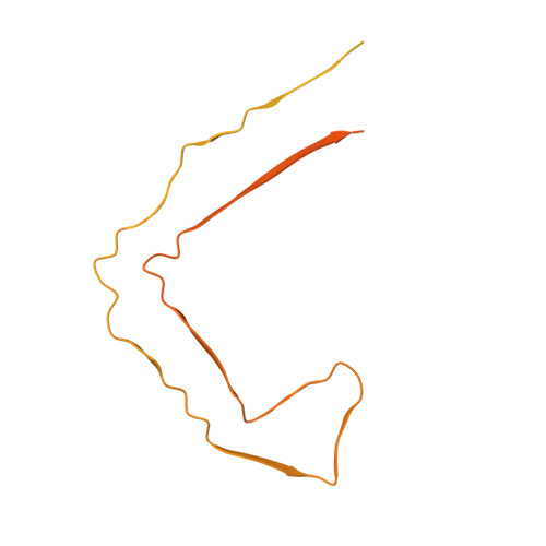
Disease-specific tau filaments assemble via polymorphic intermediates.
Lovestam, S., Li, D., Wagstaff, J.L., Kotecha, A., Kimanius, D., McLaughlin, S.H., Murzin, A.G., Freund, S.M.V., Goedert, M., Scheres, S.H.W.(2024) Nature 625: 119-125
- PubMed: 38030728
- DOI: https://doi.org/10.1038/s41586-023-06788-w
- Primary Citation of Related Structures:
8PPO, 8Q27, 8Q2J, 8Q2K, 8Q2L, 8Q7F, 8Q7L, 8Q7M, 8Q7P, 8Q7T, 8Q88, 8Q8C, 8Q8D, 8Q8E, 8Q8F, 8Q8L, 8Q8M, 8Q8R, 8Q8S, 8Q8U, 8Q8V, 8Q8W, 8Q8X, 8Q8Y, 8Q8Z, 8Q97, 8Q98, 8Q99, 8Q9A, 8Q9B, 8Q9C, 8Q9D, 8Q9E, 8Q9F, 8Q9G, 8Q9H, 8Q9I, 8Q9J, 8Q9K, 8Q9L, 8Q9M, 8Q9O, 8QCP, 8QCR, 8QJJ - PubMed Abstract:
Intermediate species in the assembly of amyloid filaments are believed to play a central role in neurodegenerative diseases and may constitute important targets for therapeutic intervention 1,2 . However, structural information about intermediate species has been scarce and the molecular mechanisms by which amyloids assemble remain largely unknown. Here we use time-resolved cryogenic electron microscopy to study the in vitro assembly of recombinant truncated tau (amino acid residues 297-391) into paired helical filaments of Alzheimer's disease or into filaments of chronic traumatic encephalopathy 3 . We report the formation of a shared first intermediate amyloid filament, with an ordered core comprising residues 302-316. Nuclear magnetic resonance indicates that the same residues adopt rigid, β-strand-like conformations in monomeric tau. At later time points, the first intermediate amyloid disappears and we observe many different intermediate amyloid filaments, with structures that depend on the reaction conditions. At the end of both assembly reactions, most intermediate amyloids disappear and filaments with the same ordered cores as those from human brains remain. Our results provide structural insights into the processes of primary and secondary nucleation of amyloid assembly, with implications for the design of new therapies.
- MRC Laboratory of Molecular Biology, Cambridge, UK.
Organizational Affiliation:


