8Q7O | pdb_00008q7o

Crystal structure of the FZD3 cysteine-rich domain in complex with a nanobody (14478)


Experimental Data Snapshot

  • Method: X-RAY DIFFRACTION
  • Resolution: 1.76 Å
  • R-Value Free: 
    0.222 (Depositor), 0.225 (DCC) 
  • R-Value Work: 
    0.217 (Depositor), 0.219 (DCC) 
  • R-Value Observed: 
    0.217 (Depositor) 

Starting Model: experimental
View more details

wwPDB Validation 3D Report Full Report

Validation slider image for 8Q7O

Ligand Structure Quality Assessment 


This is version 1.2 of the entry. See complete history

Literature

Structural insights into Frizzled3 through nanobody modulators.

Hillier, J.Zhao, Y.Carrique, L.Malinauskas, T.Ruza, R.R.Chang, T.H.Yi, G.Duyvesteyn, H.M.E.Yu, J.Lu, W.Pardon, E.Steyaert, J.Zhu, Y.Ni, T.Jones, E.Y.

(2024) Nat Commun 15: 7228-7228

  • DOI: https://doi.org/10.1038/s41467-024-51451-1
  • Primary Citation Related Structures: 
    8Q7O, 8QW4

  • PubMed Abstract: 

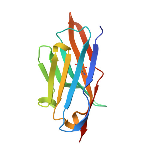
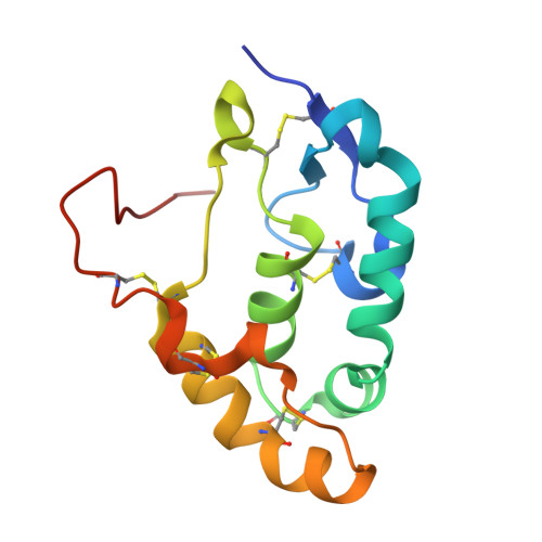
    The Wnt receptor Frizzled3 (FZD3) is important for brain axonal development and cancer progression. We report structures of FZD3 in complex with extracellular and intracellular binding nanobodies (Nb). The crystal structure of Nb8 in complex with the FZD3 cysteine-rich domain (CRD) reveals that the nanobody binds at the base of the lipid-binding groove and can compete with Wnt5a. Nb8 fused with the Dickkopf-1 C-terminal domain behaves as a FZD3-specific Wnt surrogate, activating β-catenin signalling. The cryo-EM structure of FZD3 in complex with Nb9 reveals partially resolved density for the CRD, which exhibits positional flexibility, and a transmembrane conformation that resembles active GPCRs. Nb9 binds to the cytoplasmic region of FZD3 at the putative Dishevelled (DVL) or G protein-binding site, competes with DVL binding, and inhibits GαS coupling. In combination, our FZD3 structures with nanobody modulators map extracellular and intracellular interaction surfaces of functional, and potentially therapeutic, relevance.


  • Organizational Affiliation
    • Division of Structural Biology, Wellcome Centre for Human Genetics, University of Oxford, Oxford, UK.

Macromolecule Content 

  • Total Structure Weight: 56.79 kDa 
  • Atom Count: 4,081 
  • Modeled Residue Count: 486 
  • Deposited Residue Count: 496 
  • Unique protein chains: 2

Macromolecules

Find similar proteins by:|  3D Structure
Entity ID: 1
MoleculeChains  Sequence LengthOrganismDetailsImage
Nanobody 14478
A, B
124Lama glamaMutation(s): 0 
Find similar proteins by:|  3D Structure
Entity ID: 2
MoleculeChains  Sequence LengthOrganismDetailsImage
Frizzled-3
C, D
124Homo sapiensMutation(s): 0 
Gene Names: FZD3
UniProt & NIH Common Fund Data Resources
Find proteins for Q9NPG1 (Homo sapiens)
Explore Q9NPG1 
Go to UniProtKB:  Q9NPG1
PHAROS:  Q9NPG1
GTEx:  ENSG00000104290 
Entity Groups
Sequence Clusters30% Identity50% Identity70% Identity90% Identity95% Identity100% Identity
UniProt GroupQ9NPG1
Glycosylation
Glycosylation Sites: 1Go to GlyGen: Q9NPG1-1
Sequence Annotations
Expand
Reference Sequence

Small Molecules

Ligands 5 Unique
IDChains Name / Formula / InChI Key2D Diagram3D Interactions
NAG

Query on NAG


Download:Ideal Coordinates CCD File
H [auth C],
M [auth D]
2-acetamido-2-deoxy-beta-D-glucopyranose
C8 H15 N O6
OVRNDRQMDRJTHS-FMDGEEDCSA-N
CIT

Query on CIT


Download:Ideal Coordinates CCD File
G [auth B]CITRIC ACID
C6 H8 O7
KRKNYBCHXYNGOX-UHFFFAOYSA-N
SO4

Query on SO4


Download:Ideal Coordinates CCD File
I [auth C],
N [auth D],
O [auth D]
SULFATE ION
O4 S
QAOWNCQODCNURD-UHFFFAOYSA-L
GOL

Query on GOL


Download:Ideal Coordinates CCD File
F [auth B],
J [auth C],
L [auth D]
GLYCEROL
C3 H8 O3
PEDCQBHIVMGVHV-UHFFFAOYSA-N
DMS

Query on DMS


Download:Ideal Coordinates CCD File
E [auth A],
K [auth C]
DIMETHYL SULFOXIDE
C2 H6 O S
IAZDPXIOMUYVGZ-UHFFFAOYSA-N

Experimental Data & Validation

Experimental Data

  • Method: X-RAY DIFFRACTION
  • Resolution: 1.76 Å
  • R-Value Free:  0.222 (Depositor), 0.225 (DCC) 
  • R-Value Work:  0.217 (Depositor), 0.219 (DCC) 
  • R-Value Observed: 0.217 (Depositor) 
Space Group: C 2 2 21
Unit Cell:
Length ( Å )Angle ( ˚ )
a = 53.659α = 90
b = 147.475β = 90
c = 229.727γ = 90
Software Package:
Software NamePurpose
PHENIXrefinement

Structure Validation

View Full Validation Report


Ligand Structure Quality Assessment 


Entry History 

& Funding Information

Deposition Data


Funding OrganizationLocationGrant Number
Cancer Research UKUnited Kingdom--

Revision History  (Full details and data files)

  • Version 1.0: 2024-08-28
    Type: Initial release
  • Version 1.1: 2024-09-04
    Changes: Database references
  • Version 1.2: 2024-11-13
    Changes: Structure summary