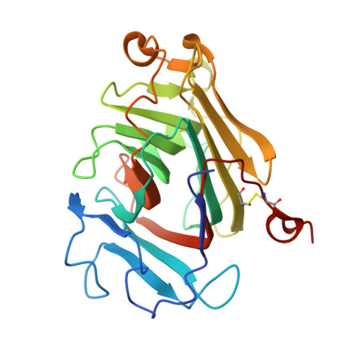
8Q29

TtX122A - A domain of unknown function from the Teredinibacter turnerae protein TERTU_3803


Experimental Data Snapshot

  • Method: X-RAY DIFFRACTION
  • Resolution: 1.50 Å
  • R-Value Free: 0.156 
  • R-Value Work: 0.112 

wwPDB Validation   3D Report Full Report


This is version 1.2 of the entry. See complete history


Literature

Structural dissection of two redox proteins from the shipworm symbiont Teredinibacter turnerae.

Rajagopal, B.S.Yates, N.Smith, J.Paradisi, A.Tetard-Jones, C.Willats, W.G.T.Marcus, S.Knox, J.P.Firdaus-Raih, M.Henrissat, B.Davies, G.J.Walton, P.H.Parkin, A.Hemsworth, G.R.

(2024) IUCrJ 11: 260-274

  • DOI: https://doi.org/10.1107/S2052252524001386
  • Primary Citation of Related Structures:  
    8Q1V, 8Q1W, 8Q28, 8Q29, 8Q2A

  • PubMed Abstract: 

    The discovery of lytic polysaccharide monooxygenases (LPMOs), a family of copper-dependent enzymes that play a major role in polysaccharide degradation, has revealed the importance of oxidoreductases in the biological utilization of biomass. In fungi, a range of redox proteins have been implicated as working in harness with LPMOs to bring about polysaccharide oxidation. In bacteria, less is known about the interplay between redox proteins and LPMOs, or how the interaction between the two contributes to polysaccharide degradation. We therefore set out to characterize two previously unstudied proteins from the shipworm symbiont Teredinibacter turnerae that were initially identified by the presence of carbohydrate binding domains appended to uncharacterized domains with probable redox functions. Here, X-ray crystal structures of several domains from these proteins are presented together with initial efforts to characterize their functions. The analysis suggests that the target proteins are unlikely to function as LPMO electron donors, raising new questions as to the potential redox functions that these large extracellular multi-haem-containing c-type cytochromes may perform in these bacteria.


  • Organizational Affiliation

    Astbury Centre for Structural Molecular Biology and School of Molecular and Cellular Biology, Faculty of Biological Sciences, University of Leeds, Leeds LS2 9JT, United Kingdom.


Macromolecules
Find similar proteins by:  (by identity cutoff)  |  3D Structure
Entity ID: 1
MoleculeChains Sequence LengthOrganismDetailsImage
Gluconolactonase domain protein
A, B
248Teredinibacter turneraeMutation(s): 0 
Gene Names: TERTU_3803
UniProt
Find proteins for C5BSV8 (Teredinibacter turnerae (strain ATCC 39867 / T7901))
Explore C5BSV8 
Go to UniProtKB:  C5BSV8
Entity Groups  
Sequence Clusters30% Identity50% Identity70% Identity90% Identity95% Identity100% Identity
UniProt GroupC5BSV8
Sequence Annotations
Expand
  • Reference Sequence
Small Molecules
Ligands 3 Unique
IDChains Name / Formula / InChI Key2D Diagram3D Interactions
EDO
Query on EDO

Download Ideal Coordinates CCD File 
F [auth A]
G [auth A]
K [auth B]
L [auth B]
M [auth B]
F [auth A],
G [auth A],
K [auth B],
L [auth B],
M [auth B],
N [auth B]
1,2-ETHANEDIOL
C2 H6 O2
LYCAIKOWRPUZTN-UHFFFAOYSA-N
CA
Query on CA

Download Ideal Coordinates CCD File 
C [auth A],
H [auth B]
CALCIUM ION
Ca
BHPQYMZQTOCNFJ-UHFFFAOYSA-N
MG
Query on MG

Download Ideal Coordinates CCD File 
D [auth A],
E [auth A],
I [auth B],
J [auth B]
MAGNESIUM ION
Mg
JLVVSXFLKOJNIY-UHFFFAOYSA-N
Experimental Data & Validation

Experimental Data

  • Method: X-RAY DIFFRACTION
  • Resolution: 1.50 Å
  • R-Value Free: 0.156 
  • R-Value Work: 0.112 
  • Space Group: P 1 21 1
Unit Cell:
Length ( Å )Angle ( ˚ )
a = 48.081α = 90
b = 75.167β = 107.423
c = 69.074γ = 90
Software Package:
Software NamePurpose
REFMACrefinement
Aimlessdata scaling
SHELXphasing
XDSdata reduction

Structure Validation

View Full Validation Report


Entry History & Funding Information

Deposition Data


Funding OrganizationLocationGrant Number
Biotechnology and Biological Sciences Research Council (BBSRC)United KingdomBB/N019970/1

Revision History  (Full details and data files)

  • Version 1.0: 2024-03-13
    Type: Initial release
  • Version 1.1: 2024-03-20
    Changes: Database references
  • Version 1.2: 2024-11-20
    Changes: Structure summary