


Crystal structures of Disulfide oxidoreductase/isomerase from Helicobacter hepaticus.
Roszczenko-Jasinska, P.To be published.
Experimental Data Snapshot
Starting Model: in silico
View more details
wwPDB Validation 3D Report Full Report

Entity ID: 1 | |||||
|---|---|---|---|---|---|
| Molecule | Chains | Sequence Length | Organism | Details | Image |
| Disulfide isomerase DsbG N-terminal domain-containing protein | 236 | Helicobacter hepaticus ATCC 51449 | Mutation(s): 0 Gene Names: HH_1141 | 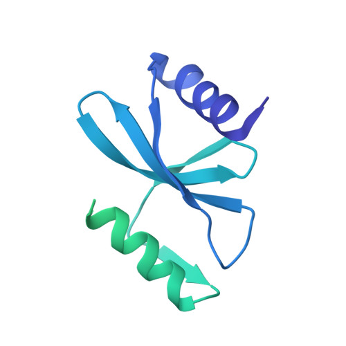 | |
UniProt | |||||
Entity Groups | |||||
| Sequence Clusters | 30% Identity50% Identity70% Identity90% Identity95% Identity100% Identity | ||||
| UniProt Group | Q7VH26 | ||||
Sequence AnnotationsExpand | |||||
Reference Sequence | |||||
| Length ( Å ) | Angle ( ˚ ) |
|---|---|
| a = 66.004 | α = 90 |
| b = 66.004 | β = 90 |
| c = 113.245 | γ = 90 |
| Software Name | Purpose |
|---|---|
| PHENIX | refinement |
| XDS | data scaling |
| PHASER | phasing |
| XDS | data reduction |
| MxCuBE | data collection |
| Funding Organization | Location | Grant Number |
|---|---|---|
| Polish National Science Centre | Poland | 2018/29/B/NZ1/00140 |


