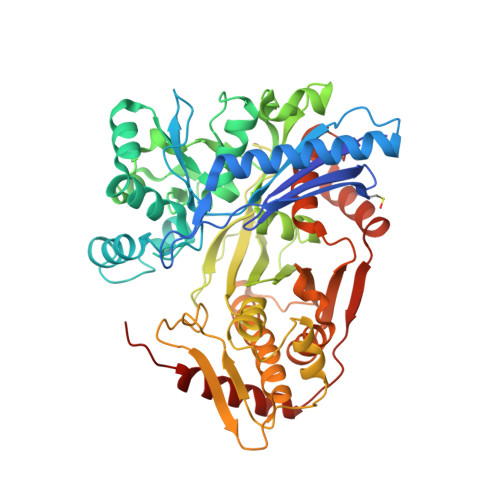
Crystal structure of glycerol kinase from Trypanosoma cruzi, a potential molecular target in Chagas disease.
Lipinski, O., Sonani, R.R., Dubin, G.(2024) Acta Crystallogr D Struct Biol 80: 629-638
- PubMed: 39052317 Search on PubMed
- DOI: https://doi.org/10.1107/S2059798324006594
- Primary Citation Related Structures:
8PRY - PubMed Abstract:
Chagas disease is a neglected tropical disease caused by the protozoan parasite Trypanosoma cruzi. It bears a significant global health burden with limited treatment options, thus calling for the development of new and effective drugs. Certain trypanosomal metabolic enzymes have been suggested to be druggable and valid for subsequent inhibition. In this study, the crystal structure of glycerol kinase from T. cruzi, a key enzyme in glycerol metabolism in this parasite, is presented. Structural analysis allowed a detailed description of the glycerol binding pocket, while comparative assessment pinpointed a potential regulatory site which may serve as a target for selective inhibition. These findings advance the understanding of glycerol metabolism in eukaryotes and provide a solid basis for the future treatment of Chagas disease.
- Malopolska Centre of Biotechnology, Jagiellonian University, Krakow, Poland.
Organizational Affiliation: