8PPG | pdb_00008ppg

Human inositol 1,4,5-trisphosphate 3-kinase A (IP3K) catalytic domain in complex with beta-D-glucopyranosyl 1,3,4,6-tetrakisphosphate/ADP/Mn after reaction


Experimental Data Snapshot

  • Method: X-RAY DIFFRACTION
  • Resolution: 1.75 Å
  • R-Value Free: 
    0.243 (Depositor), 0.246 (DCC) 
  • R-Value Work: 
    0.206 (Depositor), 0.214 (DCC) 
  • R-Value Observed: 
    0.208 (Depositor) 

Starting Model: experimental
View more details

wwPDB Validation 3D Report Full Report

Validation slider image for 8PPG

Ligand Structure Quality Assessment 


This is version 1.1 of the entry. See complete history

Literature

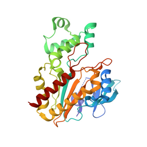
Substrate promiscuity of inositol 1,4,5-trisphosphate kinase driven by structurally-modified ligands and active site plasticity.

Marquez-Monino, M.A.Ortega-Garcia, R.Whitfield, H.Riley, A.M.Infantes, L.Garrett, S.W.Shipton, M.L.Brearley, C.A.Potter, B.V.L.Gonzalez, B.

(2024) Nat Commun 15: 1502-1502

  • DOI: https://doi.org/10.1038/s41467-024-45917-5
  • Primary Citation Related Structures: 
    8PP8, 8PP9, 8PPA, 8PPB, 8PPC, 8PPD, 8PPE, 8PPF, 8PPG, 8PPH, 8PPI, 8PPJ

  • PubMed Abstract: 

    D-myo-inositol 1,4,5-trisphosphate (InsP 3 ) is a fundamental second messenger in cellular Ca 2+ mobilization. InsP 3 3-kinase, a highly specific enzyme binding InsP 3 in just one mode, phosphorylates InsP 3 specifically at its secondary 3-hydroxyl group to generate a tetrakisphosphate. Using a chemical biology approach with both synthetised and established ligands, combining synthesis, crystallography, computational docking, HPLC and fluorescence polarization binding assays using fluorescently-tagged InsP 3 , we have surveyed the limits of InsP 3 3-kinase ligand specificity and uncovered surprisingly unforeseen biosynthetic capacity. Structurally-modified ligands exploit active site plasticity generating a helix-tilt. These facilitated uncovering of unexpected substrates phosphorylated at a surrogate extended primary hydroxyl at the inositol pseudo 3-position, applicable even to carbohydrate-based substrates. Crystallization experiments designed to allow reactions to proceed in situ facilitated unequivocal characterization of the atypical tetrakisphosphate products. In summary, we define features of InsP 3 3-kinase plasticity and substrate tolerance that may be more widely exploitable.


  • Organizational Affiliation
    • Department of Crystallography and Structural Biology, Institute of Physical-Chemistry Blas Cabrera, CSIC, Serrano 119, 28006, Madrid, Spain.

Macromolecule Content 

  • Total Structure Weight: 66.64 kDa 
  • Atom Count: 4,694 
  • Modeled Residue Count: 517 
  • Deposited Residue Count: 558 
  • Unique protein chains: 1

Macromolecules

Find similar proteins by:|  3D Structure
Entity ID: 1
MoleculeChains  Sequence LengthOrganismDetailsImage
Inositol-trisphosphate 3-kinase A
A, B
279Homo sapiensMutation(s): 0 
Gene Names: ITPKA
EC: 2.7.1.127
UniProt & NIH Common Fund Data Resources
Find proteins for P23677 (Homo sapiens)
Explore P23677 
Go to UniProtKB:  P23677
PHAROS:  P23677
GTEx:  ENSG00000137825 
Entity Groups
Sequence Clusters30% Identity50% Identity70% Identity90% Identity95% Identity100% Identity
UniProt GroupP23677
Sequence Annotations
Expand
Reference Sequence

Small Molecules

Ligands 4 Unique
IDChains Name / Formula / InChI Key2D Diagram3D Interactions
46I
(Subject of Investigation/LOI)

Query on 46I


Download:Ideal Coordinates CCD File
G [auth A],
N [auth B]
beta-D-glucopyranosyl 1,3,4,6-tetrakisphosphate
C6 H16 O18 P4
HWPKOYDCMYQLFR-UKFBFLRUSA-N
ADP
(Subject of Investigation/LOI)

Query on ADP


Download:Ideal Coordinates CCD File
F [auth A],
M [auth B]
ADENOSINE-5'-DIPHOSPHATE
C10 H15 N5 O10 P2
XTWYTFMLZFPYCI-KQYNXXCUSA-N
SO4

Query on SO4


Download:Ideal Coordinates CCD File
C [auth A]
D [auth A]
E [auth A]
J [auth A]
K [auth B]
C [auth A],
D [auth A],
E [auth A],
J [auth A],
K [auth B],
L [auth B],
P [auth B],
Q [auth B],
R [auth B]
SULFATE ION
O4 S
QAOWNCQODCNURD-UHFFFAOYSA-L
MN
(Subject of Investigation/LOI)

Query on MN


Download:Ideal Coordinates CCD File
H [auth A],
I [auth A],
O [auth B]
MANGANESE (II) ION
Mn
WAEMQWOKJMHJLA-UHFFFAOYSA-N

Experimental Data & Validation

Experimental Data

  • Method: X-RAY DIFFRACTION
  • Resolution: 1.75 Å
  • R-Value Free:  0.243 (Depositor), 0.246 (DCC) 
  • R-Value Work:  0.206 (Depositor), 0.214 (DCC) 
  • R-Value Observed: 0.208 (Depositor) 
Space Group: C 2 2 21
Unit Cell:
Length ( Å )Angle ( ˚ )
a = 71.283α = 90
b = 97.102β = 90
c = 190.755γ = 90
Software Package:
Software NamePurpose
REFMACrefinement
XDSdata reduction
XDSdata scaling
REFMACphasing
Cootmodel building
PDB_EXTRACTdata extraction

Structure Validation

View Full Validation Report


Ligand Structure Quality Assessment 


Entry History 

& Funding Information

Deposition Data


Funding OrganizationLocationGrant Number
Spanish Ministry of Science, Innovation, and UniversitiesSpainPID2020-117400GB-100
Spanish Ministry of Economy and CompetitivenessSpainBFU2017-89913-P

Revision History  (Full details and data files)

  • Version 1.0: 2024-02-28
    Type: Initial release
  • Version 1.1: 2024-03-27
    Changes: Database references