8PM1 | pdb_00008pm1

Structure of Chloroflexus aggregans flavin based fluorescent protein (CagFbFP) variant I52V A85Q


Experimental Data Snapshot

  • Method: X-RAY DIFFRACTION
  • Resolution: 1.50 Å
  • R-Value Free: 
    0.229 (Depositor), 0.227 (DCC) 
  • R-Value Work: 
    0.197 (Depositor), 0.197 (DCC) 

Starting Model: experimental
View more details

wwPDB Validation 3D Report Full Report

Validation slider image for 8PM1

Ligand Structure Quality Assessment 


This is version 1.1 of the entry. See complete history

Literature

Two distinct mechanisms of flavoprotein spectral tuning revealed by low-temperature and time-dependent spectroscopy.

Nikolaev, A.Tropina, E.V.Boldyrev, K.N.Maksimov, E.G.Borshchevskiy, V.Mishin, A.Yudenko, A.Kuzmin, A.Kuznetsova, E.Semenov, O.Remeeva, A.Gushchin, I.

(2024) Protein Sci 33: e4851-e4851

  • DOI: https://doi.org/10.1002/pro.4851
  • Primary Citation Related Structures: 
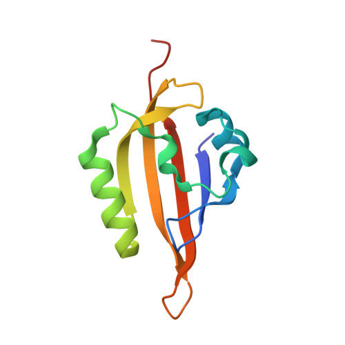
    8PKY, 8PM1

  • PubMed Abstract: 

    Flavins such as flavin mononucleotide or flavin adenine dinucleotide are bound by diverse proteins, yet have very similar spectra when in the oxidized state. Recently, we developed new variants of flavin-binding protein CagFbFP exhibiting notable blue (Q148V) or red (I52V A85Q) shifts of fluorescence emission maxima. Here, we use time-resolved and low-temperature spectroscopy to show that whereas the chromophore environment is static in Q148V, an additional protein-flavin hydrogen bond is formed upon photoexcitation in the I52V A85Q variant. Consequently, in Q148V, excitation, emission, and phosphorescence spectra are shifted, whereas in I52V A85Q, excitation and low-temperature phosphorescence spectra are relatively unchanged, while emission spectrum is altered. We also determine the x-ray structures of the two variants to reveal the flavin environment and complement the spectroscopy data. Our findings illustrate two distinct color-tuning mechanisms of flavin-binding proteins and could be helpful for the engineering of new variants with improved optical properties.


  • Organizational Affiliation
    • Research Center for Molecular Mechanisms of Aging and Age-Related Diseases, Moscow Institute of Physics and Technology, Dolgoprudny, Russia.

Macromolecule Content 

  • Total Structure Weight: 25.8 kDa 
  • Atom Count: 2,018 
  • Modeled Residue Count: 214 
  • Deposited Residue Count: 226 
  • Unique protein chains: 1

Macromolecules

Find similar proteins by:|  3D Structure
Entity ID: 1
MoleculeChains  Sequence LengthOrganismDetailsImage
histidine kinase
A, B
113Chloroflexus aggregansMutation(s): 2 
Gene Names: Cagg_3753
EC: 2.7.13.3
UniProt
Find proteins for B8GAY9 (Chloroflexus aggregans (strain MD-66 / DSM 9485))
Explore B8GAY9 
Go to UniProtKB:  B8GAY9
Entity Groups
Sequence Clusters30% Identity50% Identity70% Identity90% Identity95% Identity100% Identity
UniProt GroupB8GAY9
Sequence Annotations
Expand
Reference Sequence

Experimental Data & Validation

Experimental Data

  • Method: X-RAY DIFFRACTION
  • Resolution: 1.50 Å
  • R-Value Free:  0.229 (Depositor), 0.227 (DCC) 
  • R-Value Work:  0.197 (Depositor), 0.197 (DCC) 
Space Group: P 21 21 2
Unit Cell:
Length ( Å )Angle ( ˚ )
a = 53.817α = 90
b = 110.431β = 90
c = 39.314γ = 90
Software Package:
Software NamePurpose
REFMACrefinement
XDSdata reduction
Aimlessdata scaling
REFMACphasing

Structure Validation

View Full Validation Report


Ligand Structure Quality Assessment 


Entry History 

& Funding Information

Deposition Data


Funding OrganizationLocationGrant Number
Not funded--

Revision History  (Full details and data files)

  • Version 1.0: 2023-07-05
    Type: Initial release
  • Version 1.1: 2024-01-17
    Changes: Data collection, Database references