


Fragment-based assembly of a symmetric protein scaffold
Wouters, S.M.L., Noguchi, H., Voet, A.R.D.To be published.
Experimental Data Snapshot
wwPDB Validation 3D Report Full Report

Entity ID: 1 | |||||
|---|---|---|---|---|---|
| Molecule | Chains | Sequence Length | Organism | Details | Image |
| non-specific serine/threonine protein kinase | 86 | Mycobacterium tuberculosis | Mutation(s): 0 Gene Names: pknD_1, pknD_2, ERS007657_00042, ERS007663_00011, ERS007665_00696, ERS007720_00884, SAMEA2683035_02262 EC: 2.7.11.1 | 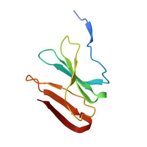 | |
UniProt | |||||
Entity Groups | |||||
| Sequence Clusters | 30% Identity50% Identity70% Identity90% Identity95% Identity100% Identity | ||||
| UniProt Group | P9WI79 | ||||
Sequence AnnotationsExpand | |||||
Reference Sequence | |||||
| Length ( Å ) | Angle ( ˚ ) |
|---|---|
| a = 95.351 | α = 90 |
| b = 95.351 | β = 90 |
| c = 69.829 | γ = 120 |
| Software Name | Purpose |
|---|---|
| PHENIX | refinement |
| Aimless | data scaling |
| XDS | data reduction |
| PHASER | phasing |
| Funding Organization | Location | Grant Number |
|---|---|---|
| Research Foundation - Flanders (FWO) | Belgium | 1S89918N |
| Research Foundation - Flanders (FWO) | Belgium | G0F9316N |
| Research Foundation - Flanders (FWO) | Belgium | G051917N |


