8PFH | pdb_00008pfh

Crystal structure of the yeast septin complex Shs1-Cdc12-Cdc3-Cdc10


Experimental Data Snapshot

  • Method: X-RAY DIFFRACTION
  • Resolution: 3.24 Å
  • R-Value Free: 
    0.290 (Depositor), 0.289 (DCC) 
  • R-Value Work: 
    0.282 (Depositor), 0.282 (DCC) 
  • R-Value Observed: 
    0.283 (Depositor) 

Starting Model: in silico
View more details

wwPDB Validation 3D Report Full Report

Validation slider image for 8PFH

Ligand Structure Quality Assessment 


This is version 1.1 of the entry. See complete history

Literature

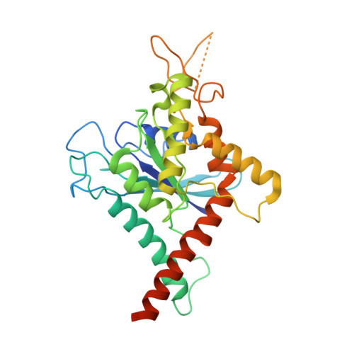
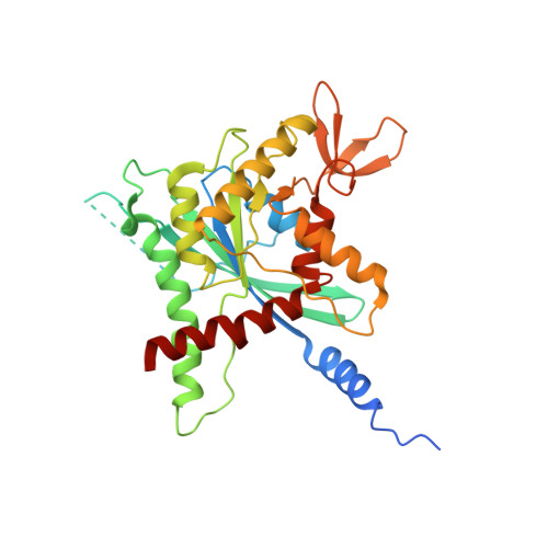
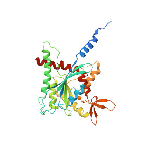
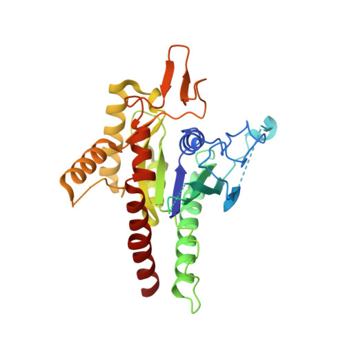
The structure of a tetrameric septin complex reveals a hydrophobic element essential for NC-interface integrity.

Grupp, B.Denkhaus, L.Gerhardt, S.Vogele, M.Johnsson, N.Gronemeyer, T.

(2024) Commun Biol 7: 48-48

  • DOI: https://doi.org/10.1038/s42003-023-05734-w
  • Primary Citation Related Structures: 
    8PFH

  • PubMed Abstract: 

    The septins of the yeast Saccharomyces cerevisiae assemble into hetero-octameric rods by alternating interactions between neighboring G-domains or N- and C-termini, respectively. These rods polymerize end to end into apolar filaments, forming a ring beneath the prospective new bud that expands during the cell cycle into an hourglass structure. The hourglass finally splits during cytokinesis into a double ring. Understanding these transitions as well as the plasticity of the higher order assemblies requires a detailed knowledge of the underlying structures. Here we present the first X-ray crystal structure of a tetrameric Shs1-Cdc12-Cdc3-Cdc10 complex at a resolution of 3.2 Å. Close inspection of the NC-interfaces of this and other septin structures reveals a conserved contact motif that is essential for NC-interface integrity of yeast and human septins in vivo and in vitro. Using the tetrameric structure in combination with AlphaFold-Multimer allowed us to propose a model of the octameric septin rod.


  • Organizational Affiliation
    • Institute of Molecular Genetics and Cell Biology, Ulm University, Ulm, Germany.

Macromolecule Content 

  • Total Structure Weight: 148.97 kDa 
  • Atom Count: 9,086 
  • Modeled Residue Count: 1,139 
  • Deposited Residue Count: 1,282 
  • Unique protein chains: 4

Macromolecules

Find similar proteins by:|  3D Structure
Entity ID: 1
MoleculeChains  Sequence LengthOrganismDetailsImage
CDC10 isoform 1300Saccharomyces cerevisiaeMutation(s): 0 
Gene Names: CDC10
UniProt
Find proteins for P25342 (Saccharomyces cerevisiae (strain ATCC 204508 / S288c))
Explore P25342 
Go to UniProtKB:  P25342
Entity Groups
Sequence Clusters30% Identity50% Identity70% Identity90% Identity95% Identity100% Identity
UniProt GroupP25342
Sequence Annotations
Expand
Reference Sequence
Find similar proteins by:|  3D Structure
Entity ID: 2
MoleculeChains  Sequence LengthOrganismDetailsImage
Cell division control protein 3332Saccharomyces cerevisiaeMutation(s): 0 
Gene Names: CDC3YLR314CL8543.7
UniProt
Find proteins for P32457 (Saccharomyces cerevisiae (strain ATCC 204508 / S288c))
Explore P32457 
Go to UniProtKB:  P32457
Entity Groups
Sequence Clusters30% Identity50% Identity70% Identity90% Identity95% Identity100% Identity
UniProt GroupP32457
Sequence Annotations
Expand
Reference Sequence
Find similar proteins by:|  3D Structure
Entity ID: 3
MoleculeChains  Sequence LengthOrganismDetailsImage
CDC12 isoform 1330Saccharomyces cerevisiaeMutation(s): 0 
Gene Names: CDC12
UniProt
Find proteins for P32468 (Saccharomyces cerevisiae (strain ATCC 204508 / S288c))
Explore P32468 
Go to UniProtKB:  P32468
Entity Groups
Sequence Clusters30% Identity50% Identity70% Identity90% Identity95% Identity100% Identity
UniProt GroupP32468
Sequence Annotations
Expand
Reference Sequence
Find similar proteins by:|  3D Structure
Entity ID: 4
MoleculeChains  Sequence LengthOrganismDetailsImage
SHS1 isoform 1320Saccharomyces cerevisiaeMutation(s): 0 
Gene Names: SHS1
UniProt
Find proteins for Q07657 (Saccharomyces cerevisiae (strain ATCC 204508 / S288c))
Explore Q07657 
Go to UniProtKB:  Q07657
Entity Groups
Sequence Clusters30% Identity50% Identity70% Identity90% Identity95% Identity100% Identity
UniProt GroupQ07657
Sequence Annotations
Expand
Reference Sequence

Experimental Data & Validation

Experimental Data

  • Method: X-RAY DIFFRACTION
  • Resolution: 3.24 Å
  • R-Value Free:  0.290 (Depositor), 0.289 (DCC) 
  • R-Value Work:  0.282 (Depositor), 0.282 (DCC) 
  • R-Value Observed: 0.283 (Depositor) 
Space Group: C 1 2 1
Unit Cell:
Length ( Å )Angle ( ˚ )
a = 258.178α = 90
b = 69.373β = 109.68
c = 92.533γ = 90
Software Package:
Software NamePurpose
PHENIXrefinement
XDSdata reduction
autoPROCdata scaling
PHASERphasing

Structure Validation

View Full Validation Report


Ligand Structure Quality Assessment 


Entry History 

& Funding Information

Deposition Data


Funding OrganizationLocationGrant Number
Not funded--

Revision History  (Full details and data files)

  • Version 1.0: 2023-12-06
    Type: Initial release
  • Version 1.1: 2024-02-21
    Changes: Database references