8PEQ | pdb_00008peq

Complex of diubiquitin-derived artificial binding protein (Affilin) variant Af2 with its target oncofetal fibronectin (fragment 7B8)


Experimental Data Snapshot

  • Method: X-RAY DIFFRACTION
  • Resolution: 2.32 Å
  • R-Value Free: 
    0.251 (Depositor), 0.251 (DCC) 
  • R-Value Work: 
    0.198 (Depositor), 0.198 (DCC) 
  • R-Value Observed: 
    0.201 (Depositor) 

Starting Models: experimental
View more details

wwPDB Validation 3D Report Full Report

Validation slider image for 8PEQ

This is version 1.1 of the entry. See complete history

Literature

Ubiquitin-derived artificial binding proteins targeting oncofetal fibronectin reveal scaffold plasticity by beta-strand slippage.

Katzschmann, A.Haupts, U.Reimann, A.Settele, F.Gloser-Braunig, M.Fiedler, E.Parthier, C.

(2024) Commun Biol 7: 907-907

  • DOI: https://doi.org/10.1038/s42003-024-06569-9
  • Primary Citation Related Structures: 
    8PEQ, 8PF0

  • PubMed Abstract: 

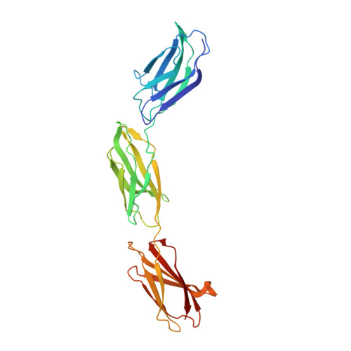
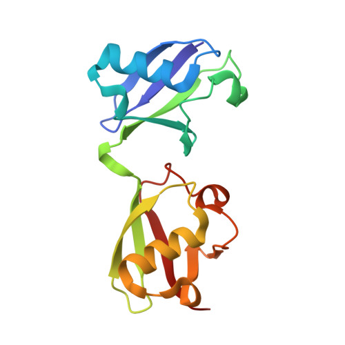
    Affilin proteins, artificial binding proteins based on the ubiquitin scaffold, have been generated by directed protein evolution to yield de-novo variants that bind the extra-domain B (EDB) of oncofetal fibronectin, an established marker of tumor neovasculature. The crystal structures of two EDB-specific Affilin variants reveal a striking structural plasticity of the ubiquitin scaffold, characterised by β-strand slippage, leading to different negative register shifts of the β5 strands. This process recruits amino acid residues from β5 towards the N-terminus to an adjacent loop region and subsequent residues into β5, respectively, remodeling the binding interface and leading to target specificity and affinity. Protein backbone alterations resulting from β-strand register shifts, as seen in the ubiquitin fold, can pose additional challenges to protein engineering as structural evidence of these events is still limited and they are difficult to predict. However, they can surface under the selection pressure of directed evolution and suggest that backbone plasticity allowing β-strand slippages can increase structural diversity, enhancing the evolutionary potential of a protein scaffold.


  • Organizational Affiliation
    • Navigo Proteins GmbH, Heinrich-Damerow-Straße 1, 06120, Halle (Saale), Germany.

Macromolecule Content 

  • Total Structure Weight: 141.91 kDa 
  • Atom Count: 10,486 
  • Modeled Residue Count: 1,278 
  • Deposited Residue Count: 1,284 
  • Unique protein chains: 2

Macromolecules

Find similar proteins by:|  3D Structure
Entity ID: 1
MoleculeChains  Sequence LengthOrganismDetailsImage
Fibronectin
A, B, C
275Homo sapiensMutation(s): 1 
Gene Names: FN1FN
UniProt & NIH Common Fund Data Resources
Find proteins for P02751 (Homo sapiens)
Explore P02751 
Go to UniProtKB:  P02751
PHAROS:  P02751
GTEx:  ENSG00000115414 
Entity Groups
Sequence Clusters30% Identity50% Identity70% Identity90% Identity95% Identity100% Identity
UniProt GroupP02751
Sequence Annotations
Expand
Reference Sequence
Find similar proteins by:|  3D Structure
Entity ID: 2
MoleculeChains  Sequence LengthOrganismDetailsImage
Affilin variant Af2D [auth J],
E [auth L],
F [auth M]
153Homo sapiensMutation(s): 0 

Experimental Data & Validation

Experimental Data

  • Method: X-RAY DIFFRACTION
  • Resolution: 2.32 Å
  • R-Value Free:  0.251 (Depositor), 0.251 (DCC) 
  • R-Value Work:  0.198 (Depositor), 0.198 (DCC) 
  • R-Value Observed: 0.201 (Depositor) 
Space Group: C 1 2 1
Unit Cell:
Length ( Å )Angle ( ˚ )
a = 168.65α = 90
b = 105.856β = 93.53
c = 78.993γ = 90
Software Package:
Software NamePurpose
PHENIXrefinement
XSCALEdata scaling
XDSdata reduction
PHASERphasing

Structure Validation

View Full Validation Report


Entry History 

& Funding Information

Deposition Data


Funding OrganizationLocationGrant Number
Not funded--

Revision History  (Full details and data files)

  • Version 1.0: 2024-06-26
    Type: Initial release
  • Version 1.1: 2024-08-14
    Changes: Database references