Helical ultrastructure of the L-ENA spore aggregation factor of a Bacillus paranthracis foodborne outbreak strain.
Sleutel, M., Zegeye, E.D., Llarena, A.K., Pradhan, B., Fislage, M., O'Sullivan, K., Van Gerven, N., Aspholm, M., Remaut, H.(2024) Nat Commun 15: 7514-7514
- PubMed: 39209852 Search on PubMedSearch on PubMed Central
- DOI: https://doi.org/10.1038/s41467-024-51804-w
- Primary Citation Related Structures:
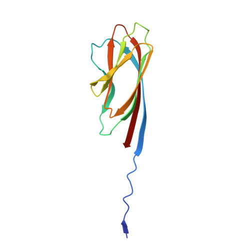
8PDZ - PubMed Abstract:
In pathogenic Bacillota, spores can form an infectious particle and can take up a central role in the environmental persistence and dissemination of disease. A poorly understood aspect of spore-mediated infection is the fibrous structures or 'endospore appendages' (ENAs) that have been seen to decorate the spores of pathogenic Bacilli and Clostridia. Current methodological approaches are opening a window on these long enigmatic structures. Using cryoID, Alphafold modelling and genetic approaches we identify a sub-class of robust ENAs in a Bacillus paranthracis foodborne outbreak strain. We demonstrate that L-ENA are encoded by a rare three-gene cluster (ena3) that contains all components for the self-assembly of ladder-like protein nanofibers of stacked heptameric rings, their anchoring to the exosporium, and their termination in a trimeric 'ruffle' made of a complement C1Q-like BclA paralogue. The role of ENA fibers in spore-spore interaction and the distribution of L-ENA operon as mobile genetic elements in B. cereus s.l. strains suggest that L-ENA fibers may increase the survival, spread and virulence of these strains.
- Structural Biology Brussels, Vrije Universiteit Brussel, Brussels, Belgium. Mike.Sleutel@vub.be.
Organizational Affiliation: