8P2W

Structure of human SIT1 (focussed map / refinement)


Experimental Data Snapshot

  • Method: ELECTRON MICROSCOPY
  • Resolution: 3.76 Å
  • Aggregation State: PARTICLE 
  • Reconstruction Method: SINGLE PARTICLE 

Starting Model: experimental
View more details

wwPDB Validation   3D Report Full Report


This is version 1.1 of the entry. See complete history


Literature

Structure and function of the SIT1 proline transporter in complex with the COVID-19 receptor ACE2.

Li, H.Z.Pike, A.C.W.Lotsaris, I.Chi, G.Hansen, J.S.Lee, S.C.Rodstrom, K.E.J.Bushell, S.R.Speedman, D.Evans, A.Wang, D.He, D.Shrestha, L.Nasrallah, C.Burgess-Brown, N.A.Vandenberg, R.J.Dafforn, T.R.Carpenter, E.P.Sauer, D.B.

(2024) Nat Commun 15: 5503-5503

  • DOI: https://doi.org/10.1038/s41467-024-48921-x
  • Primary Citation of Related Structures:  
    8P2W, 8P2X, 8P2Y, 8P2Z, 8P30, 8P31

  • PubMed Abstract: 

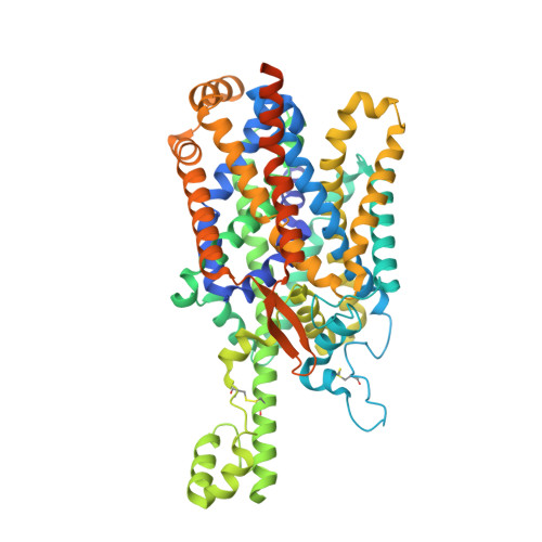
    Proline is widely known as the only proteogenic amino acid with a secondary amine. In addition to its crucial role in protein structure, the secondary amino acid modulates neurotransmission and regulates the kinetics of signaling proteins. To understand the structural basis of proline import, we solved the structure of the proline transporter SIT1 in complex with the COVID-19 viral receptor ACE2 by cryo-electron microscopy. The structure of pipecolate-bound SIT1 reveals the specific sequence requirements for proline transport in the SLC6 family and how this protein excludes amino acids with extended side chains. By comparing apo and substrate-bound SIT1 states, we also identify the structural changes that link substrate release and opening of the cytoplasmic gate and provide an explanation for how a missense mutation in the transporter causes iminoglycinuria.


  • Organizational Affiliation

    Centre for Medicines Discovery, Nuffield Department of Medicine, University of Oxford, Oxford, UK.


Macromolecules
Find similar proteins by:  (by identity cutoff)  |  3D Structure
Entity ID: 1
MoleculeChains Sequence LengthOrganismDetailsImage
Processed angiotensin-converting enzyme 2812Homo sapiensMutation(s): 0 
Gene Names: ACE2UNQ868/PRO1885
Membrane Entity: Yes 
UniProt & NIH Common Fund Data Resources
Find proteins for Q9BYF1 (Homo sapiens)
Explore Q9BYF1 
Go to UniProtKB:  Q9BYF1
GTEx:  ENSG00000130234 
Entity Groups  
Sequence Clusters30% Identity50% Identity70% Identity90% Identity95% Identity100% Identity
UniProt GroupQ9BYF1
Sequence Annotations
Expand
  • Reference Sequence
Find similar proteins by:  (by identity cutoff)  |  3D Structure
Entity ID: 2
MoleculeChains Sequence LengthOrganismDetailsImage
Sodium- and chloride-dependent transporter XTRP3B [auth C]641Homo sapiensMutation(s): 0 
Gene Names: SLC6A20SIT1XT3XTRP3
Membrane Entity: Yes 
UniProt & NIH Common Fund Data Resources
Find proteins for Q9NP91 (Homo sapiens)
Explore Q9NP91 
Go to UniProtKB:  Q9NP91
GTEx:  ENSG00000163817 
Entity Groups  
Sequence Clusters30% Identity50% Identity70% Identity90% Identity95% Identity100% Identity
UniProt GroupQ9NP91
Glycosylation
Glycosylation Sites: 2Go to GlyGen: Q9NP91-1
Sequence Annotations
Expand
  • Reference Sequence
Small Molecules
Ligands 1 Unique
IDChains Name / Formula / InChI Key2D Diagram3D Interactions
NAG
Query on NAG

Download Ideal Coordinates CCD File 
C,
D [auth C]
2-acetamido-2-deoxy-beta-D-glucopyranose
C8 H15 N O6
OVRNDRQMDRJTHS-FMDGEEDCSA-N
Experimental Data & Validation

Experimental Data

  • Method: ELECTRON MICROSCOPY
  • Resolution: 3.76 Å
  • Aggregation State: PARTICLE 
  • Reconstruction Method: SINGLE PARTICLE 
EM Software:
TaskSoftware PackageVersion
RECONSTRUCTIONcryoSPARC3.3.1
MODEL REFINEMENTPHENIX1.20_4459

Structure Validation

View Full Validation Report


Entry History & Funding Information

Deposition Data


Funding OrganizationLocationGrant Number
UK Research and Innovation (UKRI)United KingdomBB/V018051/1

Revision History  (Full details and data files)

  • Version 1.0: 2024-06-12
    Type: Initial release
  • Version 1.1: 2024-07-10
    Changes: Data collection, Database references