Stabilization of the open conformation omicron f insulin-regulated aminopeptidase by a novel substrate-selective small-molecule inhibitor.
Mpakali, A., Georgaki, G., Buson, A., Findlay, A.D., Foot, J.S., Mauvais, F.X., van Endert, P., Giastas, P., Hamprecht, D.W., Stratikos, E.(2024) Protein Sci 33: e5151-e5151
- PubMed: 39167040 Search on PubMedSearch on PubMed Central
- DOI: https://doi.org/10.1002/pro.5151
- Primary Citation Related Structures:
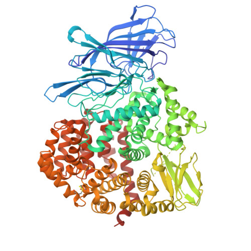
8P0I - PubMed Abstract:
Insulin-regulated aminopeptidase (IRAP) is an enzyme with important biological functions and the target of drug-discovery efforts. We combined in silico screening with a medicinal chemistry optimization campaign to discover a nanomolar inhibitor of IRAP based on a pyrazolylpyrimidine scaffold. This compound displays an excellent selectivity profile versus homologous aminopeptidases, and kinetic analysis suggests it utilizes an uncompetitive mechanism of action when inhibiting the cleavage of a typical dipeptidic substrate. Surprisingly, the compound is a poor inhibitor of the processing of the physiological cyclic peptide substrate oxytocin and a 10mer antigenic epitope precursor but displays a biphasic inhibition profile for the trimming of a 9mer antigenic peptide. While the compound reduces IRAP-dependent cross-presentation of an 8mer epitope in a cellular assay, it fails to block in vitro trimming of select epitope precursors. To gain insight into the mechanism and basis of this unusual selectivity for this inhibitor, we solved the crystal structure of its complex with IRAP. The structure indicated direct zinc(II) engagement by the pyrazolylpyrimidine scaffold and revealed that the compound binds to an open conformation of the enzyme in a pose that should block the conformational transition to the enzymatically active closed conformation previously observed for other low-molecular-weight inhibitors. This compound constitutes the first IRAP inhibitor targeting the active site that utilizes a conformation-specific mechanism of action, provides insight into the intricacies of the IRAP catalytic cycle, and highlights a novel approach to regulating IRAP activity by blocking its conformational rearrangements.
- National Centre for Scientific Research Demokritos, Athens, Greece.
Organizational Affiliation: