8OS1 | pdb_00008os1

X-ray structure of the Peroxisomal Targeting Signal 1 (PTS1) of Trypanosoma Cruzi PEX5 in complex with the PTS1 peptide


Experimental Data Snapshot

  • Method: X-RAY DIFFRACTION
  • Resolution: 2.10 Å
  • R-Value Free: 
    0.253 (Depositor), 0.253 (DCC) 
  • R-Value Work: 
    0.193 (Depositor), 0.193 (DCC) 
  • R-Value Observed: 
    0.195 (Depositor) 

Starting Model: experimental
View more details

wwPDB Validation 3D Report Full Report

Validation slider image for 8OS1

This is version 2.1 of the entry. See complete history

Literature

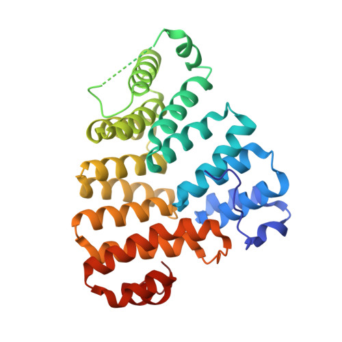
Structural dynamics of the TPR domain of the peroxisomal cargo receptor Pex5 in Trypanosoma.

Banasik, M.Napolitano, V.Blat, A.Abdulkarim, K.Plewka, J.Czaplewski, C.Gieldon, A.Kozak, M.Wladyka, B.Popowicz, G.Dubin, G.

(2024) Int J Biol Macromol 280: 135510-135510

  • DOI: https://doi.org/10.1016/j.ijbiomac.2024.135510
  • Primary Citation Related Structures: 
    8OS1, 9F8W

  • PubMed Abstract: 

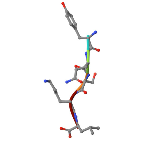
    Peroxisomal protein import has been identified as a valid target in trypanosomiases, an important health threat in Central and South America. The importomer is built of multiple peroxins (Pex) and structural characterization of these proteins facilitates rational inhibitor development. We report crystal structures of the Trypanosoma brucei and T. cruzi tetratricopeptide repeat domain (TPR) of the cytoplasmic peroxisomal targeting signal 1 (PTS1) receptor Pex5. The structure of the TPR domain of TbPex5 represents an apo-form of the receptor which, together with the previously determined structure of the complex of TbPex5 TPR and PTS1 demonstrate significant receptor dynamics associated with signal peptide recognition. The structure of the complex of TPR domain of TcPex5 with PTS1 provided in this study details the molecular interactions that guide signal peptide recognition at the atomic level in the pathogenic species currently perceived as the most relevant among Trypanosoma. Small - angle X - ray scattering (SAXS) data obtained in solution supports the crystallographic findings on the compaction of the TPR domains of TbPex5 and TcPex5 upon interaction with the cargo.


  • Organizational Affiliation
    • Malopolska Centre of Biotechnology, Jagiellonian University, Gronostajowa 7a, 30-387 Krakow, Poland.

Macromolecule Content 

  • Total Structure Weight: 75.06 kDa 
  • Atom Count: 5,252 
  • Modeled Residue Count: 615 
  • Deposited Residue Count: 656 
  • Unique protein chains: 3

Macromolecules

Find similar proteins by:|  3D Structure
Entity ID: 1
MoleculeChains  Sequence LengthOrganismDetailsImage
Peroxisome targeting signal 1 receptorA [auth B]323Trypanosoma cruziMutation(s): 0 
Gene Names: ECC02_006719
UniProt
Find proteins for Q4DPQ9 (Trypanosoma cruzi (strain CL Brener))
Explore Q4DPQ9 
Go to UniProtKB:  Q4DPQ9
Entity Groups
Sequence Clusters30% Identity50% Identity70% Identity90% Identity95% Identity100% Identity
UniProt GroupQ4DPQ9
Sequence Annotations
Expand
Reference Sequence
Find similar proteins by:|  3D Structure
Entity ID: 2
MoleculeChains  Sequence LengthOrganismDetailsImage
Peroxisome targeting signal 1 receptorB [auth A]323Trypanosoma cruziMutation(s): 0 
Gene Names: ECC02_006719
UniProt
Find proteins for Q4DPQ9 (Trypanosoma cruzi (strain CL Brener))
Explore Q4DPQ9 
Go to UniProtKB:  Q4DPQ9
Entity Groups
Sequence Clusters30% Identity50% Identity70% Identity90% Identity95% Identity100% Identity
UniProt GroupQ4DPQ9
Sequence Annotations
Expand
Reference Sequence
Find similar proteins by:  Sequence   |   3D Structure  
Entity ID: 3
MoleculeChains  Sequence LengthOrganismDetailsImage
Peroxisomal targeting signal 1 (PTS1)C [auth D],
D [auth C]
5Trypanosoma cruziMutation(s): 0 

Small Molecules

Ligands 4 Unique
IDChains Name / Formula / InChI Key2D Diagram3D Interactions
TRS

Query on TRS


Download:Ideal Coordinates CCD File
R [auth A]2-AMINO-2-HYDROXYMETHYL-PROPANE-1,3-DIOL
C4 H12 N O3
LENZDBCJOHFCAS-UHFFFAOYSA-O
PEG

Query on PEG


Download:Ideal Coordinates CCD File
F [auth B]DI(HYDROXYETHYL)ETHER
C4 H10 O3
MTHSVFCYNBDYFN-UHFFFAOYSA-N
EDO

Query on EDO


Download:Ideal Coordinates CCD File
E [auth B]
G [auth B]
H [auth B]
J [auth B]
K [auth B]
E [auth B],
G [auth B],
H [auth B],
J [auth B],
K [auth B],
L [auth B],
M [auth B],
N [auth A],
O [auth A],
P [auth A],
Q [auth A],
S [auth A],
T [auth A],
U [auth A]
1,2-ETHANEDIOL
C2 H6 O2
LYCAIKOWRPUZTN-UHFFFAOYSA-N
MG

Query on MG


Download:Ideal Coordinates CCD File
I [auth B]MAGNESIUM ION
Mg
JLVVSXFLKOJNIY-UHFFFAOYSA-N
Modified Residues  1 Unique
IDChains TypeFormula2D DiagramParent
CME
Query on CME
A [auth B]L-PEPTIDE LINKINGC5 H11 N O3 S2CYS

Experimental Data & Validation

Experimental Data

  • Method: X-RAY DIFFRACTION
  • Resolution: 2.10 Å
  • R-Value Free:  0.253 (Depositor), 0.253 (DCC) 
  • R-Value Work:  0.193 (Depositor), 0.193 (DCC) 
  • R-Value Observed: 0.195 (Depositor) 
Space Group: C 1 2 1
Unit Cell:
Length ( Å )Angle ( ˚ )
a = 129.331α = 90
b = 69.936β = 100.05
c = 71.106γ = 90
Software Package:
Software NamePurpose
REFMACrefinement
XDSdata reduction
AutoProcessdata scaling
PHASERphasing

Structure Validation

View Full Validation Report


Entry History 

& Funding Information

Deposition Data


Funding OrganizationLocationGrant Number
Accelerated Early staGe drug dIScovery (AEGIS)European Union--
Polish National Science CentrePoland2020/39/B/NZ1/01551

Revision History  (Full details and data files)

  • Version 1.0: 2024-10-02
    Type: Initial release
  • Version 2.0: 2024-10-16
    Changes: Atomic model, Data collection, Database references, Derived calculations, Non-polymer description, Polymer sequence, Source and taxonomy, Structure summary
  • Version 2.1: 2025-08-20
    Changes: Author supporting evidence