8OQU

Structure of Mycobacterium tuberculosis beta-oxidation trifunctional enzyme in complex with Fragment-M-92


Experimental Data Snapshot

  • Method: X-RAY DIFFRACTION
  • Resolution: 2.89 Å
  • R-Value Free: 0.217 
  • R-Value Work: 0.180 
  • R-Value Observed: 0.182 

Starting Model: experimental
View more details

wwPDB Validation   3D Report Full Report


Ligand Structure Quality Assessment 


This is version 2.0 of the entry. See complete history


Literature

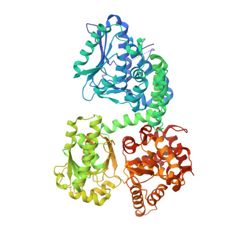
Crystallographic fragment-binding studies of the Mycobacterium tuberculosis trifunctional enzyme suggest binding pockets for the tails of the acyl-CoA substrates at its active sites and a potential substrate-channeling path between them.

Dalwani, S.Metz, A.Huschmann, F.U.Weiss, M.S.Wierenga, R.K.Venkatesan, R.

(2024) Acta Crystallogr D Struct Biol 

  • DOI: https://doi.org/10.1107/S2059798324006557
  • Primary Citation of Related Structures:  
    8OPU, 8OPV, 8OPW, 8OPX, 8OPY, 8OQL, 8OQM, 8OQN, 8OQO, 8OQP, 8OQQ, 8OQR, 8OQS, 8OQT, 8OQU, 8OQV, 8PF8

  • PubMed Abstract: 

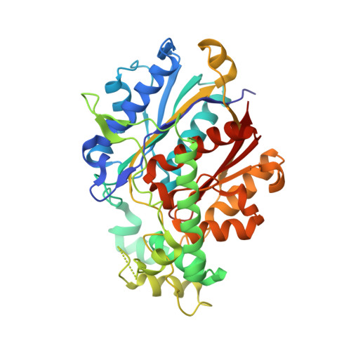
    The Mycobacterium tuberculosis trifunctional enzyme (MtTFE) is an α 2 β 2 tetrameric enzyme in which the α-chain harbors the 2E-enoyl-CoA hydratase (ECH) and 3S-hydroxyacyl-CoA dehydrogenase (HAD) active sites, and the β-chain provides the 3-ketoacyl-CoA thiolase (KAT) active site. Linear, medium-chain and long-chain 2E-enoyl-CoA molecules are the preferred substrates of MtTFE. Previous crystallographic binding and modeling studies identified binding sites for the acyl-CoA substrates at the three active sites, as well as the NAD binding pocket at the HAD active site. These studies also identified three additional CoA binding sites on the surface of MtTFE that are different from the active sites. It has been proposed that one of these additional sites could be of functional relevance for the substrate channeling (by surface crawling) of reaction intermediates between the three active sites. Here, 226 fragments were screened in a crystallographic fragment-binding study of MtTFE crystals, resulting in the structures of 16 MtTFE-fragment complexes. Analysis of the 121 fragment-binding events shows that the ECH active site is the `binding hotspot' for the tested fragments, with 41 binding events. The mode of binding of the fragments bound at the active sites provides additional insight into how the long-chain acyl moiety of the substrates can be accommodated at their proposed binding pockets. In addition, the 20 fragment-binding events between the active sites identify potential transient binding sites of reaction intermediates relevant to the possible channeling of substrates between these active sites. These results provide a basis for further studies to understand the functional relevance of the latter binding sites and to identify substrates for which channeling is crucial.


  • Organizational Affiliation

    Faculty of Biochemistry and Molecular Medicine, University of Oulu, Oulu, Finland.


Macromolecules
Find similar proteins by:  (by identity cutoff)  |  3D Structure
Entity ID: 1
MoleculeChains Sequence LengthOrganismDetailsImage
3-hydroxyacyl-CoA dehydrogenase
A, B
736Mycobacterium tuberculosis H37RvMutation(s): 0 
Gene Names: fadBRv0860
EC: 1.1.1.35
UniProt
Find proteins for O53872 (Mycobacterium tuberculosis (strain ATCC 25618 / H37Rv))
Explore O53872 
Go to UniProtKB:  O53872
Entity Groups  
Sequence Clusters30% Identity50% Identity70% Identity90% Identity95% Identity100% Identity
UniProt GroupO53872
Sequence Annotations
Expand
  • Reference Sequence
Find similar proteins by:  (by identity cutoff)  |  3D Structure
Entity ID: 2
MoleculeChains Sequence LengthOrganismDetailsImage
Putative acyltransferase Rv0859
C, D
403Mycobacterium tuberculosis H37RvMutation(s): 0 
Gene Names: fadARv0859
EC: 2.3.1
UniProt
Find proteins for O53871 (Mycobacterium tuberculosis (strain ATCC 25618 / H37Rv))
Explore O53871 
Go to UniProtKB:  O53871
Entity Groups  
Sequence Clusters30% Identity50% Identity70% Identity90% Identity95% Identity100% Identity
UniProt GroupO53871
Sequence Annotations
Expand
  • Reference Sequence
Small Molecules
Ligands 3 Unique
IDChains Name / Formula / InChI Key2D Diagram3D Interactions
VXN (Subject of Investigation/LOI)
Query on VXN

Download Ideal Coordinates CCD File 
M [auth A]
N [auth A]
O [auth A]
V [auth B]
W [auth B]
M [auth A],
N [auth A],
O [auth A],
V [auth B],
W [auth B],
X [auth B]
4-chloranylbenzenesulfonic acid
C6 H5 Cl O3 S
RJWBTWIBUIGANW-UHFFFAOYSA-N
SO4
Query on SO4

Download Ideal Coordinates CCD File 
AA [auth C]
BA [auth C]
CA [auth C]
DA [auth C]
E [auth A]
AA [auth C],
BA [auth C],
CA [auth C],
DA [auth C],
E [auth A],
EA [auth C],
F [auth A],
FA [auth D],
G [auth A],
GA [auth D],
H [auth A],
HA [auth D],
I [auth A],
IA [auth D],
JA [auth D],
P [auth B],
Q [auth B],
R [auth B],
S [auth B],
Y [auth C],
Z [auth C]
SULFATE ION
O4 S
QAOWNCQODCNURD-UHFFFAOYSA-L
GOL
Query on GOL

Download Ideal Coordinates CCD File 
J [auth A],
K [auth A],
L [auth A],
T [auth B],
U [auth B]
GLYCEROL
C3 H8 O3
PEDCQBHIVMGVHV-UHFFFAOYSA-N
Experimental Data & Validation

Experimental Data

  • Method: X-RAY DIFFRACTION
  • Resolution: 2.89 Å
  • R-Value Free: 0.217 
  • R-Value Work: 0.180 
  • R-Value Observed: 0.182 
  • Space Group: C 1 2 1
Unit Cell:
Length ( Å )Angle ( ˚ )
a = 249.462α = 90
b = 134.892β = 110.468
c = 119.476γ = 90
Software Package:
Software NamePurpose
PHENIXrefinement
XDSdata reduction
STARANISOdata scaling
PHASERphasing
Cootmodel building

Structure Validation

View Full Validation Report


Ligand Structure Quality Assessment 


Entry History & Funding Information

Deposition Data


Funding OrganizationLocationGrant Number
Academy of FinlandFinland293369
Academy of FinlandFinland289024
Academy of FinlandFinland319194

Revision History  (Full details and data files)

  • Version 1.0: 2024-01-24
    Type: Initial release
  • Version 2.0: 2024-07-24
    Type: Coordinate replacement
    Reason: Model completeness
    Changes: Advisory, Atomic model, Data collection, Database references, Derived calculations, Refinement description, Structure summary