8OG3 | pdb_00008og3

E. coli NfsB triple mutant T41L/N71S/F124T bound to citrate


Experimental Data Snapshot

  • Method: X-RAY DIFFRACTION
  • Resolution: 2.09 Å
  • R-Value Free: 
    0.218 (Depositor), 0.218 (DCC) 
  • R-Value Work: 
    0.165 (Depositor), 0.165 (DCC) 
  • R-Value Observed: 
    0.167 (Depositor) 

Starting Model: experimental
View more details

wwPDB Validation 3D Report Full Report

Validation slider image for 8OG3

Ligand Structure Quality Assessment 


This is version 1.1 of the entry. See complete history

Literature

Structure and Dynamics of Three Escherichia coli NfsB Nitro-Reductase Mutants Selected for Enhanced Activity with the Cancer Prodrug CB1954.

Day, M.A.Christofferson, A.J.Anderson, J.L.R.Vass, S.O.Evans, A.Searle, P.F.White, S.A.Hyde, E.I.

(2023) Int J Mol Sci 24

  • DOI: https://doi.org/10.3390/ijms24065987
  • Primary Citation Related Structures: 
    8C5E, 8C5F, 8C5P, 8CCV, 8CJ0, 8OG3

  • PubMed Abstract: 

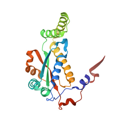
    Escherichia coli NfsB has been studied extensively for its potential for cancer gene therapy by reducing the prodrug CB1954 to a cytotoxic derivative. We have previously made several mutants with enhanced activity for the prodrug and characterised their activity in vitro and in vivo. Here, we determine the X-ray structure of our most active triple and double mutants to date, T41Q/N71S/F124T and T41L/N71S. The two mutant proteins have lower redox potentials than wild-type NfsB, and the mutations have lowered activity with NADH so that, in contrast to the wild-type enzyme, the reduction of the enzyme by NADH, rather than the reaction with CB1954, has a slower maximum rate. The structure of the triple mutant shows the interaction between Q41 and T124, explaining the synergy between these two mutations. Based on these structures, we selected mutants with even higher activity. The most active one contains T41Q/N71S/F124T/M127V, in which the additional M127V mutation enlarges a small channel to the active site. Molecular dynamics simulations show that the mutations or reduction of the FMN cofactors of the protein has little effect on its dynamics and that the largest backbone fluctuations occur at residues that flank the active site, contributing towards its broad substrate range.


  • Organizational Affiliation
    • School of Biosciences, University of Birmingham, Edgbaston, Birmingham B15 2TT, UK.

Macromolecule Content 

  • Total Structure Weight: 49 kDa 
  • Atom Count: 3,799 
  • Modeled Residue Count: 432 
  • Deposited Residue Count: 432 
  • Unique protein chains: 1

Macromolecules

Find similar proteins by:|  3D Structure
Entity ID: 1
MoleculeChains  Sequence LengthOrganismDetailsImage
Oxygen-insensitive NAD(P)H nitroreductase
A, B
216Escherichia coliMutation(s): 3 
Gene Names: nfsBdprAnfnBnfsIntrb0578JW0567
EC: 1 (PDB Primary Data), 1.5.1.34 (PDB Primary Data)
UniProt
Find proteins for P38489 (Escherichia coli (strain K12))
Explore P38489 
Go to UniProtKB:  P38489
Entity Groups
Sequence Clusters30% Identity50% Identity70% Identity90% Identity95% Identity100% Identity
UniProt GroupP38489
Sequence Annotations
Expand
Reference Sequence

Small Molecules

Ligands 3 Unique
IDChains Name / Formula / InChI Key2D Diagram3D Interactions
FMN

Query on FMN


Download:Ideal Coordinates CCD File
C [auth A],
G [auth B]
FLAVIN MONONUCLEOTIDE
C17 H21 N4 O9 P
FVTCRASFADXXNN-SCRDCRAPSA-N
CIT
(Subject of Investigation/LOI)

Query on CIT


Download:Ideal Coordinates CCD File
D [auth A],
I [auth B]
CITRIC ACID
C6 H8 O7
KRKNYBCHXYNGOX-UHFFFAOYSA-N
EDO

Query on EDO


Download:Ideal Coordinates CCD File
E [auth A],
F [auth A],
H [auth B]
1,2-ETHANEDIOL
C2 H6 O2
LYCAIKOWRPUZTN-UHFFFAOYSA-N

Experimental Data & Validation

Experimental Data

  • Method: X-RAY DIFFRACTION
  • Resolution: 2.09 Å
  • R-Value Free:  0.218 (Depositor), 0.218 (DCC) 
  • R-Value Work:  0.165 (Depositor), 0.165 (DCC) 
  • R-Value Observed: 0.167 (Depositor) 
Space Group: P 41 21 2
Unit Cell:
Length ( Å )Angle ( ˚ )
a = 57.263α = 90
b = 57.263β = 90
c = 263.827γ = 90
Software Package:
Software NamePurpose
REFMACrefinement
MOSFLMdata reduction
SCALAdata scaling
PHASERphasing

Structure Validation

View Full Validation Report


Ligand Structure Quality Assessment 


Entry History 

& Funding Information

Deposition Data


Funding OrganizationLocationGrant Number
Medical Research Council (MRC, United Kingdom)United Kingdom--

Revision History  (Full details and data files)

  • Version 1.0: 2023-04-19
    Type: Initial release
  • Version 1.1: 2024-06-19
    Changes: Data collection