8OFB | pdb_00008ofb

Crystal Structure of T. maritima reverse gyrase with a minimal latch, hexagonal form


Experimental Data Snapshot

  • Method: X-RAY DIFFRACTION
  • Resolution: 2.39 Å
  • R-Value Free: 
    0.272 (Depositor), 0.273 (DCC) 
  • R-Value Work: 
    0.243 (Depositor), 0.243 (DCC) 
  • R-Value Observed: 
    0.245 (Depositor) 

Starting Model: experimental
View more details

wwPDB Validation 3D Report Full Report

Validation slider image for 8OFB

This is version 1.3 of the entry. See complete history

Literature

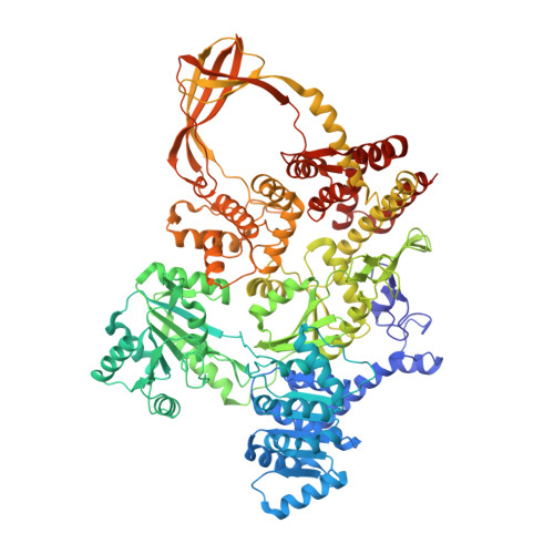
Structure of reverse gyrase with a minimal latch that supports ATP-dependent positive supercoiling without specific interactions with the topoisomerase domain.

Mhaindarkar, V.P.Rasche, R.Kummel, D.Rudolph, M.G.Klostermeier, D.

(2023) Acta Crystallogr D Struct Biol 79: 498-507

  • DOI: https://doi.org/10.1107/S2059798323002565
  • Primary Citation Related Structures: 
    7FSE, 7FSF, 8OFB

  • PubMed Abstract: 

    Reverse gyrase is the only topoisomerase that introduces positive supercoils into DNA in an ATP-dependent reaction. Positive DNA supercoiling becomes possible through the functional cooperation of the N-terminal helicase domain of reverse gyrase with its C-terminal type IA topoisomerase domain. This cooperation is mediated by a reverse-gyrase-specific insertion into the helicase domain termed the `latch'. The latch consists of a globular domain inserted at the top of a β-bulge loop that connects this globular part to the helicase domain. While the globular domain shows little conservation in sequence and length and is dispensable for DNA supercoiling, the β-bulge loop is required for supercoiling activity. It has previously been shown that the β-bulge loop constitutes a minimal latch that couples ATP-dependent processes in the helicase domain to DNA processing by the topoisomerase domain. Here, the crystal structure of Thermotoga maritima reverse gyrase with such a β-bulge loop as a minimal latch is reported. It is shown that the β-bulge loop supports ATP-dependent DNA supercoiling of reverse gyrase without engaging in specific interactions with the topoisomerase domain. When only a small latch or no latch is present, a helix in the nearby helicase domain of T. maritima reverse gyrase partially unfolds. Comparison of the sequences and predicted structures of latch regions in other reverse gyrases shows that neither sequence nor structure are decisive factors for latch functionality; instead, the decisive factors are likely to be electrostatics and plain steric bulk.


  • Organizational Affiliation
    • Institute for Physical Chemistry, University of Muenster, Corrensstrasse 30, 48149 Muenster, Germany.

Macromolecule Content 

  • Total Structure Weight: 122.31 kDa 
  • Atom Count: 8,679 
  • Modeled Residue Count: 1,040 
  • Deposited Residue Count: 1,043 
  • Unique protein chains: 1

Macromolecules

Find similar proteins by:|  3D Structure
Entity ID: 1
MoleculeChains  Sequence LengthOrganismDetailsImage
Reverse gyrase1,043Thermotoga maritima MSB8Mutation(s): 0 
Gene Names: rgytopRTM_0173
EC: 3.6.4.12 (PDB Primary Data), 5.6.2.2 (PDB Primary Data), 5.6.2 (UniProt)
UniProt
Find proteins for O51934 (Thermotoga maritima (strain ATCC 43589 / DSM 3109 / JCM 10099 / NBRC 100826 / MSB8))
Explore O51934 
Go to UniProtKB:  O51934
Entity Groups
Sequence Clusters30% Identity50% Identity70% Identity90% Identity95% Identity100% Identity
UniProt GroupO51934
Sequence Annotations
Expand
Reference Sequence

Small Molecules

Ligands 4 Unique
IDChains Name / Formula / InChI Key2D Diagram3D Interactions
P6G

Query on P6G


Download:Ideal Coordinates CCD File
B [auth A]HEXAETHYLENE GLYCOL
C12 H26 O7
IIRDTKBZINWQAW-UHFFFAOYSA-N
SO4

Query on SO4


Download:Ideal Coordinates CCD File
E [auth A],
F [auth A],
G [auth A],
H [auth A],
I [auth A]
SULFATE ION
O4 S
QAOWNCQODCNURD-UHFFFAOYSA-L
ZN

Query on ZN


Download:Ideal Coordinates CCD File
C [auth A],
D [auth A]
ZINC ION
Zn
PTFCDOFLOPIGGS-UHFFFAOYSA-N
CL

Query on CL


Download:Ideal Coordinates CCD File
J [auth A],
K [auth A]
CHLORIDE ION
Cl
VEXZGXHMUGYJMC-UHFFFAOYSA-M

Experimental Data & Validation

Experimental Data

  • Method: X-RAY DIFFRACTION
  • Resolution: 2.39 Å
  • R-Value Free:  0.272 (Depositor), 0.273 (DCC) 
  • R-Value Work:  0.243 (Depositor), 0.243 (DCC) 
  • R-Value Observed: 0.245 (Depositor) 
Space Group: P 61 2 2
Unit Cell:
Length ( Å )Angle ( ˚ )
a = 90.833α = 90
b = 90.833β = 90
c = 571.878γ = 120
Software Package:
Software NamePurpose
Aimlessdata scaling
PHENIXrefinement
PDB_EXTRACTdata extraction
xia2data reduction
PHASERphasing

Structure Validation

View Full Validation Report


Entry History 

& Funding Information

Deposition Data


Funding OrganizationLocationGrant Number
F. Hoffmann-La Roche LTDSwitzerland--

Revision History  (Full details and data files)

  • Version 1.0: 2023-04-26
    Type: Initial release
  • Version 1.1: 2023-05-31
    Changes: Database references
  • Version 1.2: 2023-06-07
    Changes: Database references
  • Version 1.3: 2024-06-19
    Changes: Data collection