8OE1 | pdb_00008oe1

Structure of P167S BlaC from Mycobacterium tuberculosis at pH 5


Experimental Data Snapshot

  • Method: X-RAY DIFFRACTION
  • Resolution: 1.90 Å
  • R-Value Free: 
    0.213 (Depositor), 0.218 (DCC) 
  • R-Value Work: 
    0.172 (Depositor), 0.183 (DCC) 
  • R-Value Observed: 
    0.174 (Depositor) 

Starting Model: experimental
View more details

wwPDB Validation 3D Report Full Report

Validation slider image for 8OE1

This is version 1.3 of the entry. See complete history

Literature

Enhanced activity against a third-generation cephalosporin by destabilization of the active site of a class A beta-lactamase.

Sun, J.Chikunova, A.Boyle, A.L.Voskamp, P.Timmer, M.Ubbink, M.

(2023) Int J Biol Macromol 250: 126160-126160

  • DOI: https://doi.org/10.1016/j.ijbiomac.2023.126160
  • Primary Citation Related Structures: 
    8OE1, 8OE5

  • PubMed Abstract: 

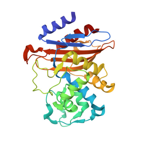
    The β-lactamase BlaC conveys resistance to a broad spectrum of β-lactam antibiotics to its host Mycobacterium tuberculosis but poorly hydrolyzes third-generation cephalosporins, such as ceftazidime. Variants of other β-lactamases have been reported to gain activity against ceftazidime at the cost of the native activity. To understand this trade-off, laboratory evolution was performed, screening for enhanced ceftazidime activity. The variant BlaC Pro167Ser shows faster breakdown of ceftazidime, poor hydrolysis of ampicillin and only moderately reduced activity against nitrocefin. NMR spectroscopy, crystallography and kinetic assays demonstrate that the resting state of BlaC P167S exists in an open and a closed state. The open state is more active in the hydrolysis of ceftazidime. In this state the catalytic residue Glu166, generally believed to be involved in the activation of the water molecule required for deacylation, is rotated away from the active site, suggesting it plays no role in the hydrolysis of ceftazidime. In the closed state, deacylation of the BlaC-ceftazidime adduct is slow, while hydrolysis of nitrocefin, which requires the presence of Glu166 in the active site, is barely affected, providing a structural explanation for the trade-off in activities.


  • Organizational Affiliation
    • Macromolecular Biochemistry, Leiden Institute of Chemistry, Leiden University, Einsteinweg 55, 2333 CC Leiden, the Netherlands.

Macromolecule Content 

  • Total Structure Weight: 57.08 kDa 
  • Atom Count: 4,229 
  • Modeled Residue Count: 530 
  • Deposited Residue Count: 530 
  • Unique protein chains: 1

Macromolecules

Find similar proteins by:|  3D Structure
Entity ID: 1
MoleculeChains  Sequence LengthOrganismDetailsImage
Beta-lactamaseA,
B [auth C]
265Mycobacterium tuberculosisMutation(s): 1 
Gene Names: blaCblaARv2068cMTCY49.07c
EC: 3.5.2.6
UniProt
Find proteins for P9WKD3 (Mycobacterium tuberculosis (strain ATCC 25618 / H37Rv))
Explore P9WKD3 
Go to UniProtKB:  P9WKD3
Entity Groups
Sequence Clusters30% Identity50% Identity70% Identity90% Identity95% Identity100% Identity
UniProt GroupP9WKD3
Sequence Annotations
Expand
Reference Sequence

Experimental Data & Validation

Experimental Data

  • Method: X-RAY DIFFRACTION
  • Resolution: 1.90 Å
  • R-Value Free:  0.213 (Depositor), 0.218 (DCC) 
  • R-Value Work:  0.172 (Depositor), 0.183 (DCC) 
  • R-Value Observed: 0.174 (Depositor) 
Space Group: P 1
Unit Cell:
Length ( Å )Angle ( ˚ )
a = 40.6α = 90.18
b = 54.178β = 111.1
c = 56.036γ = 91.78
Software Package:
Software NamePurpose
REFMACrefinement
DIALSdata reduction
Aimlessdata scaling
MOLREPphasing

Structure Validation

View Full Validation Report


Entry History 

& Funding Information

Deposition Data


Funding OrganizationLocationGrant Number
Chinese Scholarship CouncilChina--

Revision History  (Full details and data files)

  • Version 1.0: 2023-08-23
    Type: Initial release
  • Version 1.1: 2023-08-30
    Changes: Database references
  • Version 1.2: 2024-01-24
    Changes: Database references, Source and taxonomy, Structure summary
  • Version 1.3: 2024-10-02
    Changes: Data collection