8KI0 | pdb_00008ki0

Crystal structure of the hemophore HasA from Pseudomonas protegens Pf-5 capturing Fe-tetraphenylporphyrin


Experimental Data Snapshot

  • Method: X-RAY DIFFRACTION
  • Resolution: 1.45 Å
  • R-Value Free: 
    0.152 (Depositor), 0.158 (DCC) 
  • R-Value Work: 
    0.119 (Depositor), 0.126 (DCC) 
  • R-Value Observed: 
    0.121 (Depositor) 

Starting Model: in silico
View more details

wwPDB Validation 3D Report Full Report

Validation slider image for 8KI0

Ligand Structure Quality Assessment 


This is version 1.1 of the entry. See complete history

Literature

Heme-substituted protein assembly bridged by synthetic porphyrin: achieving controlled configuration while maintaining rotational freedom.

Inaba, H.Shisaka, Y.Ariyasu, S.Sakakibara, E.Ueda, G.Aiba, Y.Shimizu, N.Sugimoto, H.Shoji, O.

(2024) RSC Adv 14: 8829-8836

  • DOI: https://doi.org/10.1039/d4ra01042f
  • Primary Citation Related Structures: 
    8KI0, 8KI1

  • PubMed Abstract: 

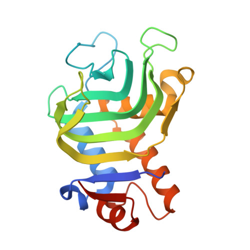
    The use of biological host-guest interactions, specifically the binding of hemoprotein to heme, has attracted significant research interest in the design of artificial protein assemblies. However, because of the inherent flexibility of the propionic acid group of heme, it is difficult to control the positioning and orientation of the protein unit and to construct well-ordered structures. Herein, we report a heme-substituted protein dimer composed of the native hemoprotein HasA, which accommodates a tetraphenylporphyrin bearing an additional metal coordination site. The specific binding of the tetraphenylporphyrin with an additional metal coordination site that protrudes in a fixed direction confines the configuration of the dimer structure to a defined bent form. The small-angle X-ray scattering profile shows the dimer structure with a bent form and suggests dynamic rotational behavior while keeping its bent-core structure, resembling a bevel gear. This unique dimer structure demonstrates that the design of heme-substituted protein assemblies can be expanded to protein assemblies while maintaining the rotational freedom of the individual protein units.


  • Organizational Affiliation
    • Department of Chemistry, School of Science, Nagoya University Furo-cho, Chikusa-ku Nagoya Aichi 464-0802 Japan shoji.osami.w3@f.mail.nagoya-u.ac.jp.

Macromolecule Content 

  • Total Structure Weight: 40.17 kDa 
  • Atom Count: 3,146 
  • Modeled Residue Count: 359 
  • Deposited Residue Count: 370 
  • Unique protein chains: 1

Macromolecules

Find similar proteins by:|  3D Structure
Entity ID: 1
MoleculeChains  Sequence LengthOrganismDetailsImage
Heme acquisition protein HasAp
A, B
185Pseudomonas protegens Pf-5Mutation(s): 0 
Gene Names: hasAp
UniProt
Find proteins for Q4K5N8 (Pseudomonas fluorescens (strain ATCC BAA-477 / NRRL B-23932 / Pf-5))
Explore Q4K5N8 
Go to UniProtKB:  Q4K5N8
Entity Groups
Sequence Clusters30% Identity50% Identity70% Identity90% Identity95% Identity100% Identity
UniProt GroupQ4K5N8
Sequence Annotations
Expand
Reference Sequence

Small Molecules

Ligands 4 Unique
IDChains Name / Formula / InChI Key2D Diagram3D Interactions
MQP
(Subject of Investigation/LOI)

Query on MQP


Download:Ideal Coordinates CCD File
C [auth A],
F [auth B]
[5,10,15,20-tetraphenylporphyrinato(2-)-kappa~4~N~21~,N~22~,N~23~,N~24~]iron
C44 H28 Fe N4
ZWYCMWUUWAFXIA-DAJBKUBHSA-N
NHE

Query on NHE


Download:Ideal Coordinates CCD File
D [auth A],
E [auth A],
G [auth B]
2-[N-CYCLOHEXYLAMINO]ETHANE SULFONIC ACID
C8 H17 N O3 S
MKWKNSIESPFAQN-UHFFFAOYSA-N
GOL

Query on GOL


Download:Ideal Coordinates CCD File
H [auth B]GLYCEROL
C3 H8 O3
PEDCQBHIVMGVHV-UHFFFAOYSA-N
NA

Query on NA


Download:Ideal Coordinates CCD File
I [auth B]SODIUM ION
Na
FKNQFGJONOIPTF-UHFFFAOYSA-N

Experimental Data & Validation

Experimental Data

  • Method: X-RAY DIFFRACTION
  • Resolution: 1.45 Å
  • R-Value Free:  0.152 (Depositor), 0.158 (DCC) 
  • R-Value Work:  0.119 (Depositor), 0.126 (DCC) 
  • R-Value Observed: 0.121 (Depositor) 
Space Group: P 1 21 1
Unit Cell:
Length ( Å )Angle ( ˚ )
a = 33.561α = 90
b = 158.717β = 97.58
c = 33.534γ = 90
Software Package:
Software NamePurpose
XDSdata scaling
REFMACrefinement
PDB_EXTRACTdata extraction
XDSdata reduction
MOLREPphasing

Structure Validation

View Full Validation Report


Ligand Structure Quality Assessment 


Entry History 

& Funding Information

Deposition Data


Funding OrganizationLocationGrant Number
Japan Society for the Promotion of Science (JSPS)JapanJP18H02084
Japan Society for the Promotion of Science (JSPS)JapanJP21H04704
Japan Society for the Promotion of Science (JSPS)JapanJP22H05129
Japan Society for the Promotion of Science (JSPS)JapanJP18J15250

Revision History  (Full details and data files)

  • Version 1.0: 2024-03-27
    Type: Initial release
  • Version 1.1: 2024-05-15
    Changes: Database references