Fusion of amyloid beta with ferritin yields an isolated oligomeric beta-sheet-rich aggregate inside the ferritin cage.
Maity, B., Kameyama, S., Tian, J., Pham, T.T., Abe, S., Chatani, E., Murata, K., Ueno, T.(2024) Biomater Sci 12: 2408-2417
- PubMed: 38511491 Search on PubMed
- DOI: https://doi.org/10.1039/d4bm00173g
- Primary Citation Related Structures:
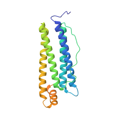
8KH2 - PubMed Abstract:
Alzheimer's disease is a severe brain condition caused by the formation of amyloid plaques composed of amyloid beta (Aβ) peptides. These peptides form oligomers, protofibrils, and fibrils before deposition into amyloid plaques. Among these intermediates, Aβ oligomers (AβOs) were found to be the most toxic and therefore an appealing target for drug development and understanding their role in the disease. However, precise isolation and characterization of AβOs have proven challenging because AβOs tend to aggregate and form heterogeneous mixtures in solution. As a solution, we genetically fused the Aβ peptide with a ferritin monomer. Such fusion allowed the encapsulation of precisely 24 Aβ peptides inside the 24-mer ferritin cage. Using high-speed atomic force microscopy (HS-AFM), we disassembled ferritin and directly visualized the Aβ core enclosed within the cage. The thioflavin-T assay (ThT) and attenuated total reflection infrared spectroscopy (ATR-IR) revealed the presence of a β-sheet structure in the encapsulated oligomeric aggregate. Gallic acid, an amyloid inhibitor, can inhibit the fluorescence of ThT bound AβOs. Our approach represents a significant advancement in the isolation and characterization of β-sheet rich AβOs and is expected to be useful for future studies of other disordered peptides such as α-synuclein and tau.
- School of Life Science and Technology, Tokyo Institute of Technology, Nagatsuta-cho, 4259, Midori-ku, Yokohama 226 8501, Japan. tueno@bio.titech.ac.jp.
Organizational Affiliation: