8KGG

LPS-bound P2Y10 in complex with G13


Experimental Data Snapshot

  • Method: ELECTRON MICROSCOPY
  • Resolution: 3.06 Å
  • Aggregation State: PARTICLE 
  • Reconstruction Method: SINGLE PARTICLE 

wwPDB Validation   3D Report Full Report


This is version 1.1 of the entry. See complete history


Literature

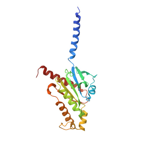
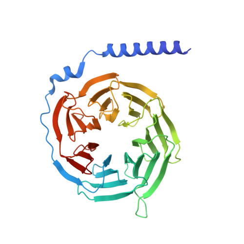
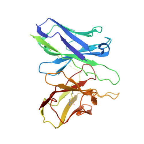
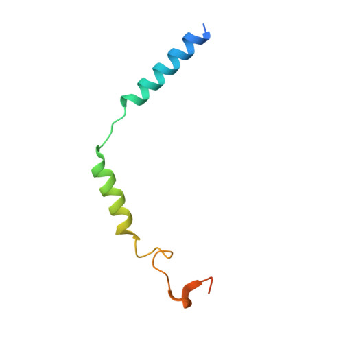
Insights into lysophosphatidylserine recognition and G alpha 12/13 -coupling specificity of P2Y10.

Yin, H.Kamakura, N.Qian, Y.Tatsumi, M.Ikuta, T.Liang, J.Xu, Z.Xia, R.Zhang, A.Guo, C.Inoue, A.He, Y.

(2024) Cell Chem Biol 

  • DOI: https://doi.org/10.1016/j.chembiol.2024.08.005
  • Primary Citation of Related Structures:  
    8KGG

  • PubMed Abstract: 

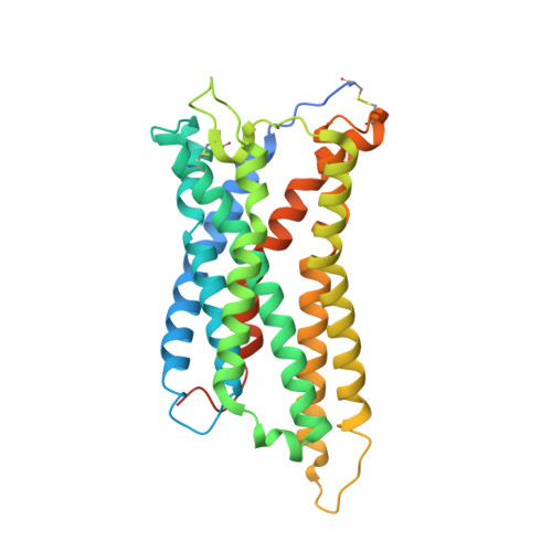
    The lysophosphatidylserine (LysoPS) receptor P2Y10, also known as LPS 2 , plays crucial roles in the regulation of immune responses and holds promise for the treatment of autoimmune diseases. Here, we report the cryoelectron microscopy (cryo-EM) structure of LysoPS-bound P2Y10 in complex with an engineered G 13 heterotrimeric protein. The structure and a mutagenesis study highlight the predominant role of a comprehensive polar network in facilitating the binding and activation of the receptor by LysoPS. This interaction pattern is preserved in GPR174, but not in GPR34. Moreover, our structural study unveils the essential interactions that underlie the Gα 13 engagement of P2Y10 and identifies key determinants for Gα 12 -vs.-Gα 13 -coupling selectivity, whose mutations selectively disrupt Gα 12 engagement while preserving the intact coupling of Gα 13 . The combined structural and functional studies provide insights into the molecular mechanisms of LysoPS recognition and Gα 12/ 13 coupling specificity.


  • Organizational Affiliation

    Laboratory of Receptor Structure and Signaling, HIT Center for Life Sciences, School of Life Science and Technology, Harbin Institute of Technology, Harbin 150001, China.


Macromolecules
Find similar proteins by:  (by identity cutoff)  |  3D Structure
Entity ID: 1
MoleculeChains Sequence LengthOrganismDetailsImage
Guanine nucleotide-binding protein G(i) subunit alpha-2,Guanine nucleotide-binding protein subunit alpha-13230Homo sapiensMutation(s): 23 
Gene Names: GNAI2GNAI2BGNA13
Membrane Entity: Yes 
UniProt & NIH Common Fund Data Resources
Find proteins for P04899 (Homo sapiens)
Explore P04899 
Go to UniProtKB:  P04899
PHAROS:  P04899
GTEx:  ENSG00000114353 
Find proteins for Q14344 (Homo sapiens)
Explore Q14344 
Go to UniProtKB:  Q14344
PHAROS:  Q14344
GTEx:  ENSG00000120063 
Entity Groups  
Sequence Clusters30% Identity50% Identity70% Identity90% Identity95% Identity100% Identity
UniProt GroupsQ14344P04899
Sequence Annotations
Expand
  • Reference Sequence
Find similar proteins by:  (by identity cutoff)  |  3D Structure
Entity ID: 2
MoleculeChains Sequence LengthOrganismDetailsImage
Guanine nucleotide-binding protein G(I)/G(S)/G(T) subunit beta-1345Homo sapiensMutation(s): 0 
Gene Names: GNB1
UniProt & NIH Common Fund Data Resources
Find proteins for P62873 (Homo sapiens)
Explore P62873 
Go to UniProtKB:  P62873
PHAROS:  P62873
GTEx:  ENSG00000078369 
Entity Groups  
Sequence Clusters30% Identity50% Identity70% Identity90% Identity95% Identity100% Identity
UniProt GroupP62873
Sequence Annotations
Expand
  • Reference Sequence
Find similar proteins by:  (by identity cutoff)  |  3D Structure
Entity ID: 3
MoleculeChains Sequence LengthOrganismDetailsImage
scFv16C [auth E]247Homo sapiensMutation(s): 0 
Entity Groups  
Sequence Clusters30% Identity50% Identity70% Identity90% Identity95% Identity100% Identity
Sequence Annotations
Expand
  • Reference Sequence
Find similar proteins by:  (by identity cutoff)  |  3D Structure
Entity ID: 4
MoleculeChains Sequence LengthOrganismDetailsImage
Guanine nucleotide-binding protein G(I)/G(S)/G(O) subunit gamma-2D [auth G]71Homo sapiensMutation(s): 0 
Gene Names: GNG2
Membrane Entity: Yes 
UniProt & NIH Common Fund Data Resources
Find proteins for P59768 (Homo sapiens)
Explore P59768 
Go to UniProtKB:  P59768
PHAROS:  P59768
GTEx:  ENSG00000186469 
Entity Groups  
Sequence Clusters30% Identity50% Identity70% Identity90% Identity95% Identity100% Identity
UniProt GroupP59768
Sequence Annotations
Expand
  • Reference Sequence
Find similar proteins by:  (by identity cutoff)  |  3D Structure
Entity ID: 5
MoleculeChains Sequence LengthOrganismDetailsImage
Putative P2Y purinoceptor 10E [auth R]339Homo sapiensMutation(s): 0 
Gene Names: P2RY10
Membrane Entity: Yes 
UniProt & NIH Common Fund Data Resources
Find proteins for O00398 (Homo sapiens)
Explore O00398 
Go to UniProtKB:  O00398
PHAROS:  O00398
GTEx:  ENSG00000078589 
Entity Groups  
Sequence Clusters30% Identity50% Identity70% Identity90% Identity95% Identity100% Identity
UniProt GroupO00398
Sequence Annotations
Expand
  • Reference Sequence
Small Molecules
Ligands 1 Unique
IDChains Name / Formula / InChI Key2D Diagram3D Interactions
WJS
Query on WJS

Download Ideal Coordinates CCD File 
F [auth R](2~{S})-2-$l^{4}-azanyl-3-[[(2~{R})-3-octadecanoyloxy-2-oxidanyl-propoxy]-oxidanyl-oxidanylidene-$l^{6}-phosphanyl]oxy-propanoic acid
C24 H20 N O9 P
JFEXCESBNOAOBB-YADHBBJMSA-N
Experimental Data & Validation

Experimental Data

  • Method: ELECTRON MICROSCOPY
  • Resolution: 3.06 Å
  • Aggregation State: PARTICLE 
  • Reconstruction Method: SINGLE PARTICLE 

Structure Validation

View Full Validation Report


Entry History & Funding Information

Deposition Data

  • Released Date: 2024-07-31 
  • Deposition Author(s): He, Y., Yin, Y.

Funding OrganizationLocationGrant Number
National Natural Science Foundation of China (NSFC)China32070048

Revision History  (Full details and data files)

  • Version 1.0: 2024-07-31
    Type: Initial release
  • Version 1.1: 2024-09-25
    Changes: Data collection, Database references