


Crystal structure of single domain antibody kt75 of human Thyroglobulin
Mistry, H., Kumarasamy, J., Kumari, S., Kulkarni, S.P., Gupta, G.D.To be published.
Experimental Data Snapshot
Starting Model: experimental
View more details
wwPDB Validation 3D Report Full Report

Entity ID: 1 | |||||
|---|---|---|---|---|---|
| Molecule | Chains | Sequence Length | Organism | Details | Image |
| single domain antibody kt75 of human Thyroglobulin | 134 | Camelus dromedarius | Mutation(s): 0 | 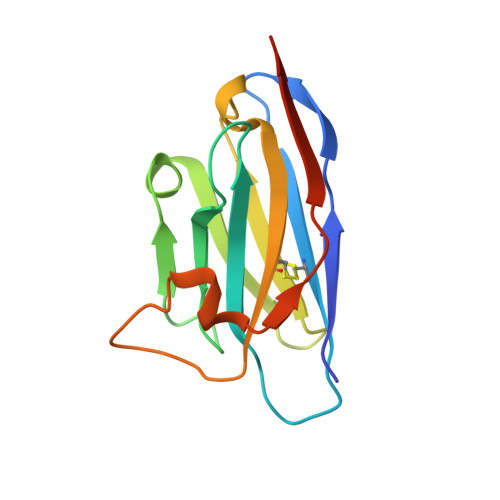 | |
| Ligands 2 Unique | |||||
|---|---|---|---|---|---|
| ID | Chains | Name / Formula / InChI Key | 2D Diagram | 3D Interactions | |
| ACT Download:Ideal Coordinates CCD File | B [auth A], C [auth A], D [auth A] | ACETATE ION C2 H3 O2 QTBSBXVTEAMEQO-UHFFFAOYSA-M |  | ||
| K Download:Ideal Coordinates CCD File | E [auth A] | POTASSIUM ION K NPYPAHLBTDXSSS-UHFFFAOYSA-N |  | ||
| Length ( Å ) | Angle ( ˚ ) |
|---|---|
| a = 38.751 | α = 90 |
| b = 47.589 | β = 90 |
| c = 64.237 | γ = 90 |
| Software Name | Purpose |
|---|---|
| REFMAC | refinement |
| PHENIX | refinement |
| XDS | data reduction |
| Aimless | data scaling |
| PHASER | phasing |
| Funding Organization | Location | Grant Number |
|---|---|---|
| Other government | India | -- |


