8JX9

rat megalin bodyA


Experimental Data Snapshot

  • Method: ELECTRON MICROSCOPY
  • Resolution: 3.80 Å
  • Aggregation State: PARTICLE 
  • Reconstruction Method: SINGLE PARTICLE 

wwPDB Validation   3D Report Full Report


This is version 1.0 of the entry. See complete history


Literature

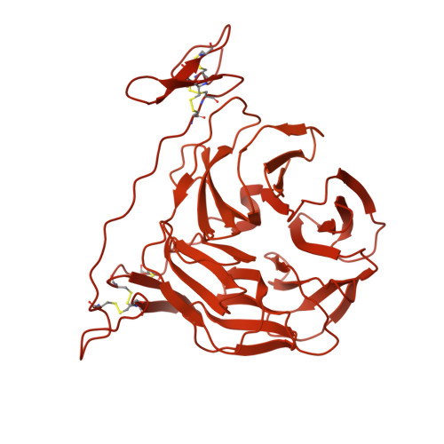
Cryo-EM structures elucidate the multiligand receptor nature of megalin.

Goto, S.Tsutsumi, A.Lee, Y.Hosojima, M.Kabasawa, H.Komochi, K.Nagatoishi, S.Takemoto, K.Tsumoto, K.Nishizawa, T.Kikkawa, M.Saito, A.

(2024) Proc Natl Acad Sci U S A 121: e2318859121-e2318859121

  • DOI: https://doi.org/10.1073/pnas.2318859121
  • Primary Citation of Related Structures:  
    8JUT, 8JUU, 8JX8, 8JX9, 8JXA, 8JXB, 8JXC, 8JXD, 8JXE, 8JXF, 8JXG, 8JXH, 8JXI, 8JXJ

  • PubMed Abstract: 

    Megalin (low-density lipoprotein receptor-related protein 2) is a giant glycoprotein of about 600 kDa, mediating the endocytosis of more than 60 ligands, including those of proteins, peptides, and drug compounds [S. Goto, M. Hosojima, H. Kabasawa, A. Saito, Int. J. Biochem. Cell Biol. 157 , 106393 (2023)]. It is expressed predominantly in renal proximal tubule epithelial cells, as well as in the brain, lungs, eyes, inner ear, thyroid gland, and placenta. Megalin is also known to mediate the endocytosis of toxic compounds, particularly those that cause renal and hearing disorders [Y. Hori et al. , J. Am. Soc. Nephrol. 28 , 1783-1791 (2017)]. Genetic megalin deficiency causes Donnai-Barrow syndrome/facio-oculo-acoustico-renal syndrome in humans. However, it is not known how megalin interacts with such a wide variety of ligands and plays pathological roles in various organs. In this study, we elucidated the dimeric architecture of megalin, purified from rat kidneys, using cryoelectron microscopy. The maps revealed the densities of endogenous ligands bound to various regions throughout the dimer, elucidating the multiligand receptor nature of megalin. We also determined the structure of megalin in complex with receptor-associated protein, a molecular chaperone for megalin. The results will facilitate further studies on the pathophysiology of megalin-dependent multiligand endocytic pathways in multiple organs and will also be useful for the development of megalin-targeted drugs for renal and hearing disorders, Alzheimer's disease [B. V. Zlokovic et al. , Proc. Natl. Acad. Sci. U.S.A. 93 , 4229-4234 (1996)], and other illnesses.


  • Organizational Affiliation

    Department of Applied Molecular Medicine, Kidney Research Center, Niigata University Graduate School of Medical and Dental Sciences, Niigata City 951-8510, Japan.


Macromolecules
Find similar proteins by:  (by identity cutoff)  |  3D Structure
Entity ID: 1
MoleculeChains Sequence LengthOrganismDetailsImage
LDL receptor related protein 2
A, B
4,660Rattus norvegicusMutation(s): 0 
UniProt
Find proteins for A0A0G2K9W7 (Rattus norvegicus)
Explore A0A0G2K9W7 
Go to UniProtKB:  A0A0G2K9W7
Entity Groups  
Sequence Clusters30% Identity50% Identity70% Identity90% Identity95% Identity100% Identity
UniProt GroupA0A0G2K9W7
Sequence Annotations
Expand
  • Reference Sequence

Find similar proteins by:  Sequence   |   3D Structure  

Entity ID: 2
MoleculeChains Sequence LengthOrganismDetailsImage
ligandC [auth O]5Rattus norvegicusMutation(s): 0 
Sequence Annotations
Expand
  • Reference Sequence
Oligosaccharides

Help

Entity ID: 3
MoleculeChains Length2D Diagram Glycosylation3D Interactions
beta-D-mannopyranose-(1-4)-2-acetamido-2-deoxy-beta-D-glucopyranose-(1-4)-2-acetamido-2-deoxy-beta-D-glucopyranoseD [auth C],
E [auth D]
3N-Glycosylation
Entity ID: 4
MoleculeChains Length2D Diagram Glycosylation3D Interactions
alpha-D-mannopyranose-(1-3)-[alpha-D-mannopyranose-(1-6)]beta-D-mannopyranose-(1-4)-2-acetamido-2-deoxy-beta-D-glucopyranose-(1-4)-2-acetamido-2-deoxy-beta-D-glucopyranoseF [auth E],
I [auth H],
J [auth I]
5N-Glycosylation
Entity ID: 5
MoleculeChains Length2D Diagram Glycosylation3D Interactions
2-acetamido-2-deoxy-beta-D-glucopyranose-(1-4)-2-acetamido-2-deoxy-beta-D-glucopyranoseG [auth F],
H [auth G]
2N-Glycosylation
Small Molecules
Ligands 3 Unique
IDChains Name / Formula / InChI Key2D Diagram3D Interactions
A2G
Query on A2G

Download Ideal Coordinates CCD File 
M [auth B],
N [auth B],
O [auth B],
P [auth B],
Q [auth B]
2-acetamido-2-deoxy-alpha-D-galactopyranose
C8 H15 N O6
OVRNDRQMDRJTHS-CBQIKETKSA-N
NAG
Query on NAG

Download Ideal Coordinates CCD File 
K [auth B],
L [auth B]
2-acetamido-2-deoxy-beta-D-glucopyranose
C8 H15 N O6
OVRNDRQMDRJTHS-FMDGEEDCSA-N
CA
Query on CA

Download Ideal Coordinates CCD File 
AA [auth B]
BA [auth B]
CA [auth B]
DA [auth B]
EA [auth B]
AA [auth B],
BA [auth B],
CA [auth B],
DA [auth B],
EA [auth B],
FA [auth B],
GA [auth B],
HA [auth B],
IA [auth B],
JA [auth B],
KA [auth B],
R [auth B],
S [auth B],
T [auth B],
U [auth B],
V [auth B],
W [auth B],
X [auth B],
Y [auth B],
Z [auth B]
CALCIUM ION
Ca
BHPQYMZQTOCNFJ-UHFFFAOYSA-N
Experimental Data & Validation

Experimental Data

  • Method: ELECTRON MICROSCOPY
  • Resolution: 3.80 Å
  • Aggregation State: PARTICLE 
  • Reconstruction Method: SINGLE PARTICLE 
EM Software:
TaskSoftware PackageVersion
MODEL REFINEMENTPHENIX
RECONSTRUCTIONRELION3.1.3

Structure Validation

View Full Validation Report


Entry History & Funding Information

Deposition Data


Funding OrganizationLocationGrant Number
Japan Society for the Promotion of Science (JSPS)Japan--

Revision History  (Full details and data files)

  • Version 1.0: 2024-06-19
    Type: Initial release