8JWJ | pdb_00008jwj

PHD Finger Protein 7 (PHF7) in complex with UBE2D2


Experimental Data Snapshot

  • Method: X-RAY DIFFRACTION
  • Resolution: 2.96 Å
  • R-Value Free: 
    0.222 (Depositor), 0.219 (DCC) 
  • R-Value Work: 
    0.194 (Depositor), 0.191 (DCC) 
  • R-Value Observed: 
    0.196 (Depositor) 

Starting Models: experimental, in silico
View more details

wwPDB Validation 3D Report Full Report

Validation slider image for 8JWJ

This is version 1.1 of the entry. See complete history

Literature

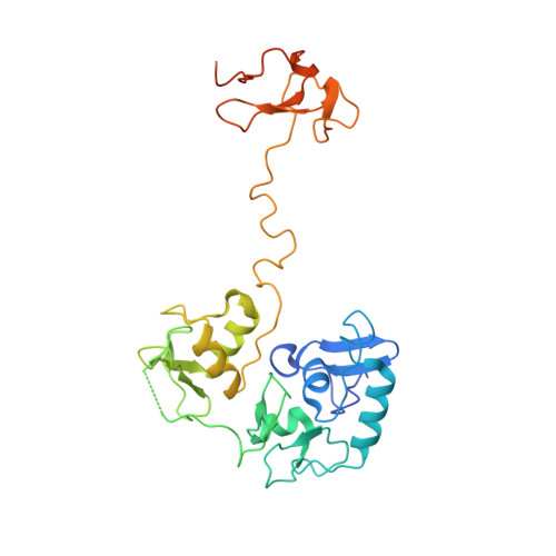
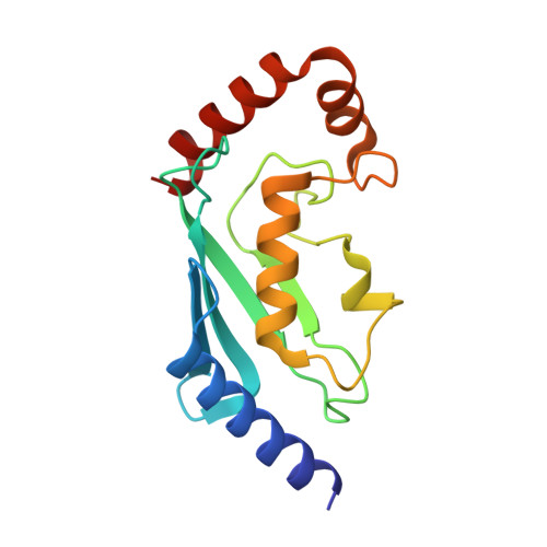
Molecular basis for PHF7-mediated ubiquitination of histone H3.

Lee, H.S.Bang, I.You, J.Jeong, T.K.Kim, C.R.Hwang, M.Kim, J.S.Baek, S.H.Song, J.J.Choi, H.J.

(2023) Genes Dev 37: 984-997

  • DOI: https://doi.org/10.1101/gad.350989.123
  • Primary Citation Related Structures: 
    8JWJ, 8JWS, 8JWU

  • PubMed Abstract: 

    The RING-type E3 ligase has been known for over two decades, yet its diverse modes of action are still the subject of active research. Plant homeodomain (PHD) finger protein 7 (PHF7) is a RING-type E3 ubiquitin ligase responsible for histone ubiquitination. PHF7 comprises three zinc finger domains: an extended PHD (ePHD), a RING domain, and a PHD. While the function of the RING domain is largely understood, the roles of the other two domains in E3 ligase activity remain elusive. Here, we present the crystal structure of PHF7 in complex with the E2 ubiquitin-conjugating enzyme (E2). Our structure shows that E2 is effectively captured between the RING domain and the C-terminal PHD, facilitating E2 recruitment through direct contact. In addition, through in vitro binding and functional assays, we demonstrate that the N-terminal ePHD recognizes the nucleosome via DNA binding, whereas the C-terminal PHD is involved in histone H3 recognition. Our results provide a molecular basis for the E3 ligase activity of PHF7 and uncover the specific yet collaborative contributions of each domain to the PHF7 ubiquitination activity.


  • Organizational Affiliation
    • Department of Biological Sciences, Seoul National University, Seoul 08826, Republic of Korea.

Macromolecule Content 

  • Total Structure Weight: 103.69 kDa 
  • Atom Count: 6,694 
  • Modeled Residue Count: 839 
  • Deposited Residue Count: 904 
  • Unique protein chains: 2

Macromolecules

Find similar proteins by:|  3D Structure
Entity ID: 1
MoleculeChains  Sequence LengthOrganismDetailsImage
PHD finger protein 7
A, C
300Mus musculusMutation(s): 0 
Gene Names: Phf7
EC: 2.3.2.27
UniProt & NIH Common Fund Data Resources
Find proteins for Q9DAG9 (Mus musculus)
Explore Q9DAG9 
Go to UniProtKB:  Q9DAG9
IMPC:  MGI:1919088
Entity Groups
Sequence Clusters30% Identity50% Identity70% Identity90% Identity95% Identity100% Identity
UniProt GroupQ9DAG9
Sequence Annotations
Expand
Reference Sequence
Find similar proteins by:|  3D Structure
Entity ID: 2
MoleculeChains  Sequence LengthOrganismDetailsImage
Ubiquitin-conjugating enzyme E2 D2
B, D
152Homo sapiensMutation(s): 2 
Gene Names: UBE2D2PUBC1UBC4UBC5BUBCH4UBCH5B
EC: 2.3.2.23 (PDB Primary Data), 2.3.2.24 (PDB Primary Data)
UniProt & NIH Common Fund Data Resources
Find proteins for P62837 (Homo sapiens)
Explore P62837 
Go to UniProtKB:  P62837
PHAROS:  P62837
GTEx:  ENSG00000131508 
Entity Groups
Sequence Clusters30% Identity50% Identity70% Identity90% Identity95% Identity100% Identity
UniProt GroupP62837
Sequence Annotations
Expand
Reference Sequence

Small Molecules

Ligands 4 Unique
IDChains Name / Formula / InChI Key2D Diagram3D Interactions
MPD

Query on MPD


Download:Ideal Coordinates CCD File
U [auth C],
V [auth C]
(4S)-2-METHYL-2,4-PENTANEDIOL
C6 H14 O2
SVTBMSDMJJWYQN-YFKPBYRVSA-N
SO4

Query on SO4


Download:Ideal Coordinates CCD File
L [auth A],
W [auth C]
SULFATE ION
O4 S
QAOWNCQODCNURD-UHFFFAOYSA-L
GOL

Query on GOL


Download:Ideal Coordinates CCD File
T [auth C],
X [auth D],
Y [auth D],
Z [auth D]
GLYCEROL
C3 H8 O3
PEDCQBHIVMGVHV-UHFFFAOYSA-N
ZN

Query on ZN


Download:Ideal Coordinates CCD File
E [auth A]
F [auth A]
G [auth A]
H [auth A]
I [auth A]
E [auth A],
F [auth A],
G [auth A],
H [auth A],
I [auth A],
J [auth A],
K [auth A],
M [auth C],
N [auth C],
O [auth C],
P [auth C],
Q [auth C],
R [auth C],
S [auth C]
ZINC ION
Zn
PTFCDOFLOPIGGS-UHFFFAOYSA-N

Experimental Data & Validation

Experimental Data

  • Method: X-RAY DIFFRACTION
  • Resolution: 2.96 Å
  • R-Value Free:  0.222 (Depositor), 0.219 (DCC) 
  • R-Value Work:  0.194 (Depositor), 0.191 (DCC) 
  • R-Value Observed: 0.196 (Depositor) 
Space Group: P 21 21 21
Unit Cell:
Length ( Å )Angle ( ˚ )
a = 102.304α = 90
b = 106.825β = 90
c = 145.759γ = 90
Software Package:
Software NamePurpose
PHENIXrefinement
XDSdata reduction
XDSdata scaling
PHENIXphasing

Structure Validation

View Full Validation Report


Entry History 

& Funding Information

Deposition Data


Funding OrganizationLocationGrant Number
National Research Foundation (NRF, Korea)Korea, Republic Of--

Revision History  (Full details and data files)

  • Version 1.0: 2023-12-20
    Type: Initial release
  • Version 1.1: 2024-01-10
    Changes: Database references