


Crystal Structure of Aspartate Semialdehyde Dehydrogenase from Porphyromonas gingivalis.
Hwang, J., Do, H., Shim, Y.S., Lee, J.H.(2023) Crystals (Basel) 13
Experimental Data Snapshot
Starting Model: in silico
View more details
(2023) Crystals (Basel) 13
Entity ID: 1 | |||||
|---|---|---|---|---|---|
| Molecule | Chains | Sequence Length | Organism | Details | Image |
| Aspartate-semialdehyde dehydrogenase | 337 | Porphyromonas gingivalis | Mutation(s): 0 Gene Names: asd, PGIN_13-1_01585 EC: 1.2.1.11 | 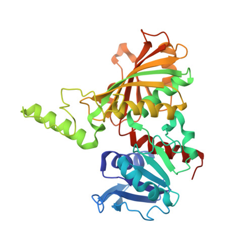 | |
| Ligands 1 Unique | |||||
|---|---|---|---|---|---|
| ID | Chains | Name / Formula / InChI Key | 2D Diagram | 3D Interactions | |
| A2P (Subject of Investigation/LOI) Download:Ideal Coordinates CCD File | C [auth A], D [auth B] | ADENOSINE-2'-5'-DIPHOSPHATE C10 H15 N5 O10 P2 AEOBEOJCBAYXBA-KQYNXXCUSA-N |  | ||
| Length ( Å ) | Angle ( ˚ ) |
|---|---|
| a = 74.99 | α = 90 |
| b = 108.22 | β = 90 |
| c = 160.4 | γ = 90 |
| Software Name | Purpose |
|---|---|
| REFMAC | refinement |
| PHENIX | refinement |
| XDS | data reduction |
| XDS | data scaling |
| MOLREP | phasing |
| Funding Organization | Location | Grant Number |
|---|---|---|
| National Research Foundation (NRF, Korea) | Korea, Republic Of | PM23030 |


