8JT1

COLLAGENASE FROM GRIMONTIA (VIBRIO) HOLLISAE 1706B COMPLEXED WITH GLY-PRO-HYP-GLY-PRO-HYP


Experimental Data Snapshot

  • Method: X-RAY DIFFRACTION
  • Resolution: 2.00 Å
  • R-Value Free: 0.214 
  • R-Value Work: 0.176 
  • R-Value Observed: 0.178 

wwPDB Validation   3D Report Full Report


Ligand Structure Quality Assessment 


This is version 1.0 of the entry. See complete history


Literature

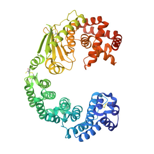
Insights into the catalytic mechanism of Grimontia hollisae collagenase through structural and mutational analyses.

Ueshima, S.Yasumoto, M.Kitagawa, Y.Akazawa, K.Takita, T.Tanaka, K.Hattori, S.Mizutani, K.Mikami, B.Yasukawa, K.

(2023) FEBS Lett 597: 2473-2483

  • DOI: https://doi.org/10.1002/1873-3468.14732
  • Primary Citation of Related Structures:  
    8JT1

  • PubMed Abstract: 

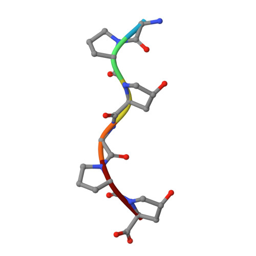
    Grimontia hollisae collagenase (Ghcol) exhibits high collagen-degrading activity. To explore its catalytic mechanism, its substrate (Gly-Pro-Hyp-Gly-Pro-Hyp, GPOGPO)-complexed crystal structure was determined at 2.0 Å resolution. A water molecule was observed near the active-site zinc ion. Since this water was not observed in the product (GPO)-complexed Ghcol, it was hypothesized that the GPOGPO-complexed Ghcol structure reflects a Michaelis complex, providing a structural basis for understanding the catalytic mechanism. Analyses of the active-site geometry and site-directed mutagenesis of the active-site tyrosine residues revealed that Glu493 and Tyr564 were essential for catalysis, suggesting that Glu493 functions as an acid and base catalyst while Tyr564 stabilizes the tetrahedral complex in the transition state. These results shed light on the catalytic mechanism of bacterial collagenase.


  • Organizational Affiliation

    Division of Food Science and Biotechnology, Graduate School of Agriculture, Kyoto University, Japan.


Macromolecules
Find similar proteins by:  (by identity cutoff)  |  3D Structure
Entity ID: 1
MoleculeChains Sequence LengthOrganismDetailsImage
Microbial collagenase
A, B
559Grimontia hollisaeMutation(s): 0 
EC: 3.4.24.3
UniProt
Find proteins for F7IZI6 (Grimontia hollisae)
Explore F7IZI6 
Go to UniProtKB:  F7IZI6
Entity Groups  
Sequence Clusters30% Identity50% Identity70% Identity90% Identity95% Identity100% Identity
UniProt GroupF7IZI6
Sequence Annotations
Expand
  • Reference Sequence

Find similar proteins by:  Sequence   |   3D Structure  

Entity ID: 2
MoleculeChains Sequence LengthOrganismDetailsImage
6-mer peptide6synthetic constructMutation(s): 0 
Sequence Annotations
Expand
  • Reference Sequence
Small Molecules
Ligands 6 Unique
IDChains Name / Formula / InChI Key2D Diagram3D Interactions
PGE (Subject of Investigation/LOI)
Query on PGE

Download Ideal Coordinates CCD File 
FB [auth A]TRIETHYLENE GLYCOL
C6 H14 O4
ZIBGPFATKBEMQZ-UHFFFAOYSA-N
TRS (Subject of Investigation/LOI)
Query on TRS

Download Ideal Coordinates CCD File 
FD [auth B]2-AMINO-2-HYDROXYMETHYL-PROPANE-1,3-DIOL
C4 H12 N O3
LENZDBCJOHFCAS-UHFFFAOYSA-O
PEG (Subject of Investigation/LOI)
Query on PEG

Download Ideal Coordinates CCD File 
ED [auth B]DI(HYDROXYETHYL)ETHER
C4 H10 O3
MTHSVFCYNBDYFN-UHFFFAOYSA-N
ZN (Subject of Investigation/LOI)
Query on ZN

Download Ideal Coordinates CCD File 
GB [auth B],
I [auth A]
ZINC ION
Zn
PTFCDOFLOPIGGS-UHFFFAOYSA-N
EDO
Query on EDO

Download Ideal Coordinates CCD File 
AA [auth A]
AB [auth A]
AC [auth B]
AD [auth B]
BA [auth A]
AA [auth A],
AB [auth A],
AC [auth B],
AD [auth B],
BA [auth A],
BB [auth A],
BC [auth B],
BD [auth B],
CA [auth A],
CB [auth A],
CC [auth B],
CD [auth B],
DA [auth A],
DB [auth A],
DC [auth B],
DD [auth B],
EA [auth A],
EB [auth A],
EC [auth B],
FA [auth A],
FC [auth B],
GA [auth A],
GC [auth B],
HA [auth A],
HC [auth B],
IA [auth A],
IC [auth B],
JA [auth A],
JC [auth B],
KA [auth A],
KC [auth B],
LA [auth A],
LB [auth B],
LC [auth B],
MA [auth A],
MB [auth B],
MC [auth B],
N [auth A],
NA [auth A],
NB [auth B],
NC [auth B],
O [auth A],
OA [auth A],
OB [auth B],
OC [auth B],
P [auth A],
PA [auth A],
PB [auth B],
PC [auth B],
Q [auth A],
QA [auth A],
QB [auth B],
QC [auth B],
R [auth A],
RA [auth A],
RB [auth B],
RC [auth B],
S [auth A],
SA [auth A],
SB [auth B],
SC [auth B],
T [auth A],
TA [auth A],
TB [auth B],
TC [auth B],
U [auth A],
UA [auth A],
UB [auth B],
UC [auth B],
V [auth A],
VA [auth A],
VB [auth B],
VC [auth B],
W [auth A],
WA [auth A],
WB [auth B],
WC [auth B],
X [auth A],
XA [auth A],
XB [auth B],
XC [auth B],
Y [auth A],
YA [auth A],
YB [auth B],
YC [auth B],
Z [auth A],
ZA [auth A],
ZB [auth B],
ZC [auth B]
1,2-ETHANEDIOL
C2 H6 O2
LYCAIKOWRPUZTN-UHFFFAOYSA-N
CA
Query on CA

Download Ideal Coordinates CCD File 
HB [auth B]
IB [auth B]
J [auth A]
JB [auth B]
K [auth A]
HB [auth B],
IB [auth B],
J [auth A],
JB [auth B],
K [auth A],
KB [auth B],
L [auth A],
M [auth A]
CALCIUM ION
Ca
BHPQYMZQTOCNFJ-UHFFFAOYSA-N
Modified Residues  1 Unique
IDChains TypeFormula2D DiagramParent
HYP
Query on HYP
C
D [auth E]
E [auth F]
F [auth G]
G [auth I]
C,
D [auth E],
E [auth F],
F [auth G],
G [auth I],
H [auth J]
L-PEPTIDE LINKINGC5 H9 N O3PRO
Experimental Data & Validation

Experimental Data

  • Method: X-RAY DIFFRACTION
  • Resolution: 2.00 Å
  • R-Value Free: 0.214 
  • R-Value Work: 0.176 
  • R-Value Observed: 0.178 
  • Space Group: C 1 2 1
Unit Cell:
Length ( Å )Angle ( ˚ )
a = 178.017α = 90
b = 74.89β = 126.02
c = 135.134γ = 90
Software Package:
Software NamePurpose
PHENIXrefinement
XDSdata reduction
XDSdata scaling
PHENIXphasing

Structure Validation

View Full Validation Report


Ligand Structure Quality Assessment 


Entry History & Funding Information

Deposition Data


Funding OrganizationLocationGrant Number
Japan Society for the Promotion of Science (JSPS)Japan22H03332

Revision History  (Full details and data files)

  • Version 1.0: 2024-02-07
    Type: Initial release