8JQ5 | pdb_00008jq5

Crystal structure of Lactobacillus rhamnosus L-rhamnose isomerase in complex with D-allulose


Experimental Data Snapshot

  • Method: X-RAY DIFFRACTION
  • Resolution: 1.73 Å
  • R-Value Free: 
    0.187 (Depositor), 0.193 (DCC) 
  • R-Value Work: 
    0.139 (Depositor), 0.151 (DCC) 
  • R-Value Observed: 
    0.142 (Depositor) 

Starting Model: experimental
View more details

wwPDB Validation 3D Report Full Report

Validation slider image for 8JQ5

Ligand Structure Quality Assessment 


This is version 1.0 of the entry. See complete history

Literature

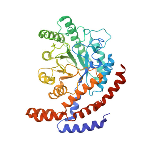
X-ray structure and characterization of a probiotic Lactobacillus rhamnosus Probio-M9 L-rhamnose isomerase.

Yoshida, H.Yamamoto, N.Kurahara, L.H.Izumori, K.Yoshihara, A.

(2024) Appl Microbiol Biotechnol 108: 249-249

  • DOI: https://doi.org/10.1007/s00253-024-13075-9
  • Primary Citation Related Structures: 
    8JQ3, 8JQ4, 8JQ5, 8JQ6

  • PubMed Abstract: 

    A recombinant L-rhamnose isomerase (L-RhI) from probiotic Lactobacillus rhamnosus Probio-M9 (L. rhamnosus Probio-M9) was expressed. L. rhamnosus Probio-M9 was isolated from human colostrum and identified as a probiotic lactic acid bacterium, which can grow using L-rhamnose. L-RhI is one of the enzymes involved in L-rhamnose metabolism and catalyzes the reversible isomerization between L-rhamnose and L-rhamnulose. Some L-RhIs were reported to catalyze isomerization not only between L-rhamnose and L-rhamnulose but also between D-allulose and D-allose, which are known as rare sugars. Those L-RhIs are attractive enzymes for rare sugar production and have the potential to be further improved by enzyme engineering; however, the known crystal structures of L-RhIs recognizing rare sugars are limited. In addition, the optimum pH levels of most reported L-RhIs are basic rather than neutral, and such a basic condition causes non-enzymatic aldose-ketose isomerization, resulting in unexpected by-products. Herein, we report the crystal structures of L. rhamnosus Probio-M9 L-RhI (LrL-RhI) in complexes with L-rhamnose, D-allulose, and D-allose, which show enzyme activity toward L-rhamnose, D-allulose, and D-allose in acidic conditions, though the activity toward D-allose was low. In the complex with L-rhamnose, L-rhamnopyranose was found in the catalytic site, showing favorable recognition for catalysis. In the complex with D-allulose, D-allulofuranose and ring-opened D-allulose were observed in the catalytic site. However, bound D-allose in the pyranose form was found in the catalytic site of the complex with D-allose, which was unfavorable for recognition, like an inhibition mode. The structure of the complex may explain the low activity toward D-allose. KEY POINTS: • Crystal structures of LrL-RhI in complexes with substrates were determined. • LrL-RhI exhibits enzyme activity toward L-rhamnose, D-allulose, and D-allose. • The LrL-RhI is active in acidic conditions.


  • Organizational Affiliation
    • Department of Basic Life Science, Faculty of Medicine, Kagawa University, 1750-1 Ikenobe, Miki-Cho, Kita-Gun, Kagawa, 761-0793, Japan. yoshida.hiromi@kagawa-u.ac.jp.

Macromolecule Content 

  • Total Structure Weight: 200.87 kDa 
  • Atom Count: 14,466 
  • Modeled Residue Count: 1,671 
  • Deposited Residue Count: 1,736 
  • Unique protein chains: 1

Macromolecules

Find similar proteins by:|  3D Structure
Entity ID: 1
MoleculeChains  Sequence LengthOrganismDetailsImage
L-rhamnose isomerase
A, B, C, D
434Lacticaseibacillus rhamnosusMutation(s): 0 
Gene Names: rhaA
EC: 5.3.1.14
UniProt
Find proteins for A0A171J5T1 (Lacticaseibacillus rhamnosus)
Explore A0A171J5T1 
Go to UniProtKB:  A0A171J5T1
Entity Groups
Sequence Clusters30% Identity50% Identity70% Identity90% Identity95% Identity100% Identity
UniProt GroupA0A171J5T1
Sequence Annotations
Expand
Reference Sequence

Small Molecules

Ligands 3 Unique
IDChains Name / Formula / InChI Key2D Diagram3D Interactions
PSV
(Subject of Investigation/LOI)

Query on PSV


Download:Ideal Coordinates CCD File
H [auth A],
O [auth C],
P [auth C],
T [auth D]
alpha-D-psicofuranose
C6 H12 O6
RFSUNEUAIZKAJO-KAZBKCHUSA-N
PSJ
(Subject of Investigation/LOI)

Query on PSJ


Download:Ideal Coordinates CCD File
G [auth A],
K [auth B],
N [auth C],
S [auth D]
D-psicose
C6 H12 O6
BJHIKXHVCXFQLS-PUFIMZNGSA-N
MN
(Subject of Investigation/LOI)

Query on MN


Download:Ideal Coordinates CCD File
E [auth A]
F [auth A]
I [auth B]
J [auth B]
L [auth C]
E [auth A],
F [auth A],
I [auth B],
J [auth B],
L [auth C],
M [auth C],
Q [auth D],
R [auth D]
MANGANESE (II) ION
Mn
WAEMQWOKJMHJLA-UHFFFAOYSA-N

Experimental Data & Validation

Experimental Data

  • Method: X-RAY DIFFRACTION
  • Resolution: 1.73 Å
  • R-Value Free:  0.187 (Depositor), 0.193 (DCC) 
  • R-Value Work:  0.139 (Depositor), 0.151 (DCC) 
  • R-Value Observed: 0.142 (Depositor) 
Space Group: P 21 21 21
Unit Cell:
Length ( Å )Angle ( ˚ )
a = 89.887α = 90
b = 139.445β = 90
c = 146.834γ = 90
Software Package:
Software NamePurpose
Aimlessdata scaling
XDSdata reduction
REFMACrefinement
MOLREPphasing

Structure Validation

View Full Validation Report


Ligand Structure Quality Assessment 


Entry History 

& Funding Information

Deposition Data


Funding OrganizationLocationGrant Number
Not funded--

Revision History  (Full details and data files)

  • Version 1.0: 2024-03-13
    Type: Initial release