8JOZ

Crystal structure of CmoM from E. coli complexed with sinefungin and cellularly expressed tRNA Ser


Experimental Data Snapshot

  • Method: X-RAY DIFFRACTION
  • Resolution: 2.22 Å
  • R-Value Free: 0.225 
  • R-Value Work: 0.199 
  • R-Value Observed: 0.200 

wwPDB Validation   3D Report Full Report


Ligand Structure Quality Assessment 


This is version 1.1 of the entry. See complete history


Literature

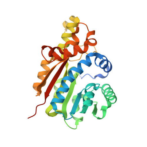
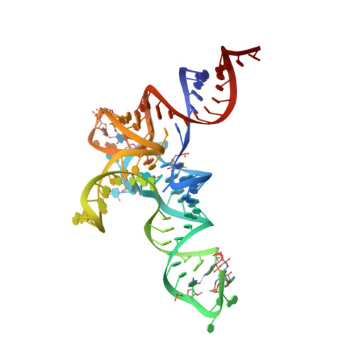
Structural basis for the selective methylation of 5-carboxymethoxyuridine in tRNA modification.

Yoo, J.Lee, J.Kim, J.

(2023) Nucleic Acids Res 51: 9432-9441

  • DOI: https://doi.org/10.1093/nar/gkad668
  • Primary Citation of Related Structures:  
    8JOZ

  • PubMed Abstract: 

    Posttranscriptional modifications of tRNA are widely conserved in all domains of life. Especially, those occurring within the anticodon often modulate translational efficiency. Derivatives of 5-hydroxyuridine are specifically found in bacterial tRNA, where 5-methoxyuridine and 5-carboxymethoxyuridine are the major species in Gram-positive and Gram-negative bacteria, respectively. In certain tRNA species, 5-carboxymethoxyuridine can be further methylated by CmoM to form the methyl ester. In this report, we present the X-ray crystal structure of Escherichia coli CmoM complexed with tRNASer1, which contains 5-carboxymethoxyuridine at the 5'-end of anticodon (the 34th position of tRNA). The 2.22 Å resolution structure of the enzyme-tRNA complex reveals that both the protein and tRNA undergo local conformational changes around the binding interface. Especially, the hypomodified uracil base is flipped out from the canonical stacked conformation enabling the specific molecular interactions with the enzyme. Moreover, the structure illustrates that the enzyme senses exclusively the anticodon arm region of the substrate tRNA and examines the presence of key determinants, 5-carboxymethoxyuridine at position 34 and guanosine at position 35, offering molecular basis for the discriminatory mechanism against non-cognate tRNAs.


  • Organizational Affiliation

    Department of Chemistry, Gwangju Institute of Science and Technology, Gwangju 61005, Korea.


Macromolecules

Find similar proteins by:  (by identity cutoff)  |  3D Structure
Entity ID: 1
MoleculeChains Sequence LengthOrganismDetailsImage
tRNA 5-carboxymethoxyuridine methyltransferase282Escherichia coli K-12Mutation(s): 0 
Gene Names: cmoMsmtAycbDb0921JW0904
EC: 2.1.1
UniProt
Find proteins for P36566 (Escherichia coli (strain K12))
Explore P36566 
Go to UniProtKB:  P36566
Entity Groups  
Sequence Clusters30% Identity50% Identity70% Identity90% Identity95% Identity100% Identity
UniProt GroupP36566
Sequence Annotations
Expand
  • Reference Sequence
Find similar nucleic acids by:  (by identity cutoff)  |  3D Structure
Entity ID: 2
MoleculeChains LengthOrganismImage
tRNA Ser (88-MER)88Escherichia coli K-12
Sequence Annotations
Expand
  • Reference Sequence
Experimental Data & Validation

Experimental Data

  • Method: X-RAY DIFFRACTION
  • Resolution: 2.22 Å
  • R-Value Free: 0.225 
  • R-Value Work: 0.199 
  • R-Value Observed: 0.200 
  • Space Group: P 61 2 2
Unit Cell:
Length ( Å )Angle ( ˚ )
a = 65.614α = 90
b = 65.614β = 90
c = 584.532γ = 120
Software Package:
Software NamePurpose
PHENIXrefinement
MxDCdata collection
XDSdata reduction
Aimlessdata scaling
MOLREPphasing
Cootmodel building
PDB_EXTRACTdata extraction

Structure Validation

View Full Validation Report


Ligand Structure Quality Assessment 


Entry History & Funding Information

Deposition Data


Funding OrganizationLocationGrant Number
National Research Foundation (NRF, Korea)Korea, Republic Of--

Revision History  (Full details and data files)

  • Version 1.0: 2023-08-30
    Type: Initial release
  • Version 1.1: 2023-10-04
    Changes: Database references