N-terminal domain truncation yielded a unique dimer of polysaccharide hydrolase with enhanced enzymatic activity, stability and calcium ion independence.
Xiang, L., Hu, X., Du, C., Wu, L., Lu, Z., Zhou, J., Zhang, G.(2024) Int J Biol Macromol 266: 131352-131352
- PubMed: 38574926 Search on PubMed
- DOI: https://doi.org/10.1016/j.ijbiomac.2024.131352
- Primary Citation Related Structures:
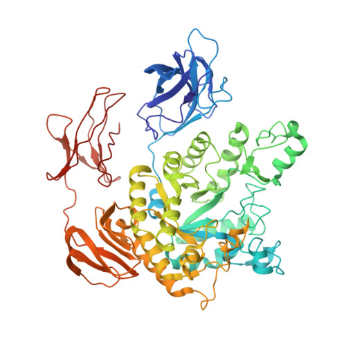
8JNX - PubMed Abstract:
Domain engineering, including domain truncation, fusion, or swapping, has become a common strategy to improve properties of enzymes, especially glycosyl hydrolases. However, there are few reports explaining the mechanism of increased activity from a protein structure perspective. Amy703 is an alkaline amylase with a unique N-terminal domain. Prior studies have shown that N-Amy, a mutant without an N-terminal domain, exhibits improved activity, stability, and calcium ion independence. In this study, we have used X-ray crystallography to determine the crystal structure of N-Amy and used AlphaFold2 to model the Amy703 structure, respectively. We further used size exclusion chromatography to show that Amy703 existed as a monomer, whereas N-Amy formed a unique dimer. It was found that the N-terminus of one monomer of N-Amy was inserted into the catalytic domain of its symmetrical subunit, resulting in the expansion of the catalytic pocket. This also significantly increased the pK a of the hydrogen donor Glu350, thereby enhancing substrate binding affinity and contributing to increased N-Amy activity. Meanwhile, two calcium ions were found to bind to N-Amy at different binding sites, which also contributed to the stability of protein. Therefore, this study provided new structural insights into the mechanisms of various glycosyl hydrolases.
- College of Life Science and Technology, Beijing University of Chemical Technology, Beijing, People's Republic of China; State Key Laboratory of Biocatalysis and Enzyme Engineering, School of Life Sciences, Hubei University, Hubei, People's Republic of China.
Organizational Affiliation: