8JMP | pdb_00008jmp

Structure of a leaf-branch compost cutinase, ICCG in complex with 1,4-butanediol terephthalate


Experimental Data Snapshot

  • Method: X-RAY DIFFRACTION
  • Resolution: 1.90 Å
  • R-Value Free: 
    0.196 (Depositor), 0.198 (DCC) 
  • R-Value Work: 
    0.154 (Depositor), 0.156 (DCC) 
  • R-Value Observed: 
    0.156 (Depositor) 

Starting Model: experimental
View more details

wwPDB Validation 3D Report Full Report

Validation slider image for 8JMP

Ligand Structure Quality Assessment 


This is version 1.2 of the entry. See complete history

Literature

Remodeling the polymer-binding cavity to improve the efficacy of PBAT-degrading enzyme.

Yang, Y.Cheng, S.Zheng, Y.Xue, T.Huang, J.W.Zhang, L.Yang, Y.Guo, R.T.Chen, C.C.

(2023) J Hazard Mater 464: 132965-132965

  • DOI: https://doi.org/10.1016/j.jhazmat.2023.132965
  • Primary Citation Related Structures: 
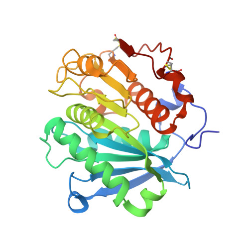
    8JMO, 8JMP

  • PubMed Abstract: 

    Poly(butylene adipate-co-terephthalate) (PBAT) is among the most widely applied synthetic polyesters that are utilized in the packaging and agricultural industries, but the accumulation of PBAT wastes has posed a great burden to ecosystems. Using renewable enzymes to decompose PBAT is an eco-friendly solution to tackle this problem. Recently, we demonstrated that cutinase is the most effective PBAT-degrading enzyme and that an engineered cutinase termed TfCut-DM could completely decompose PBAT film to terephthalate (TPA). Here, we report crystal structures of a variant of leaf compost cutinase in complex with soluble fragments of PBAT, including BTa and TaBTa. In the TaBTa complex, one TPA moiety was located at a polymer-binding site distal to the catalytic center that has never been experimentally validated. Intriguingly, the composition of the distal TPA-binding site shows higher diversity relative to the one proximal to the catalytic center in various cutinases. We thus modified the distal TPA-binding site of TfCut-DM and obtained variants that exhibit higher activity. Notably, the time needed to completely degrade the PBAT film to TPA was shortened to within 24 h by TfCut-DM Q132Y (5813 mol per mol protein). Taken together, the structural information regarding the substrate-binding behavior of PBAT-degrading enzymes could be useful guidance for direct enzyme engineering.


  • Organizational Affiliation
    • State Key Laboratory of Biocatalysis and Enzyme Engineering, Hubei Hongshan Laboratory, Hubei Collaborative Innovation Center for Green Transformation of Bio-Resources, Hubei Key Laboratory of Industrial Biotechnology, School of Life Sciences, Hubei University, 430062 Wuhan, People's Republic of China.

Macromolecule Content 

  • Total Structure Weight: 56.1 kDa 
  • Atom Count: 4,616 
  • Modeled Residue Count: 516 
  • Deposited Residue Count: 520 
  • Unique protein chains: 1

Macromolecules

Find similar proteins by:|  3D Structure
Entity ID: 1
MoleculeChains  Sequence LengthOrganismDetailsImage
Leaf-branch compost cutinase
A, B
260unidentified prokaryotic organismMutation(s): 5 
EC: 3.1.1.74 (PDB Primary Data), 3.1.1.101 (PDB Primary Data)
UniProt
Find proteins for G9BY57 (Unknown prokaryotic organism)
Explore G9BY57 
Go to UniProtKB:  G9BY57
Entity Groups
Sequence Clusters30% Identity50% Identity70% Identity90% Identity95% Identity100% Identity
UniProt GroupG9BY57
Sequence Annotations
Expand
Reference Sequence

Small Molecules

Experimental Data & Validation

Experimental Data

  • Method: X-RAY DIFFRACTION
  • Resolution: 1.90 Å
  • R-Value Free:  0.196 (Depositor), 0.198 (DCC) 
  • R-Value Work:  0.154 (Depositor), 0.156 (DCC) 
  • R-Value Observed: 0.156 (Depositor) 
Space Group: P 21 21 21
Unit Cell:
Length ( Å )Angle ( ˚ )
a = 41.19α = 90
b = 84.928β = 90
c = 147.979γ = 90
Software Package:
Software NamePurpose
PHENIXrefinement
SAINTdata reduction
SADABSdata scaling
PHENIXphasing

Structure Validation

View Full Validation Report


Ligand Structure Quality Assessment 


Entry History 

& Funding Information

Deposition Data


Funding OrganizationLocationGrant Number
National Natural Science Foundation of China (NSFC)China31870790
National Natural Science Foundation of China (NSFC)China31971205
National Natural Science Foundation of China (NSFC)China--

Revision History  (Full details and data files)

  • Version 1.0: 2023-11-29
    Type: Initial release
  • Version 1.1: 2024-11-06
    Changes: Structure summary
  • Version 1.2: 2025-07-02
    Changes: Database references