8JJ2

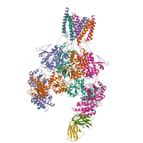
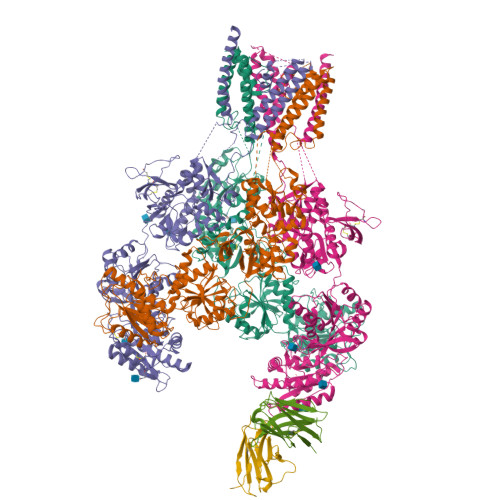
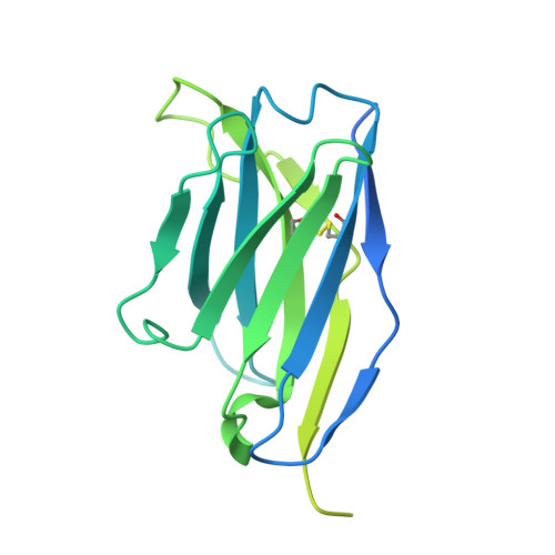
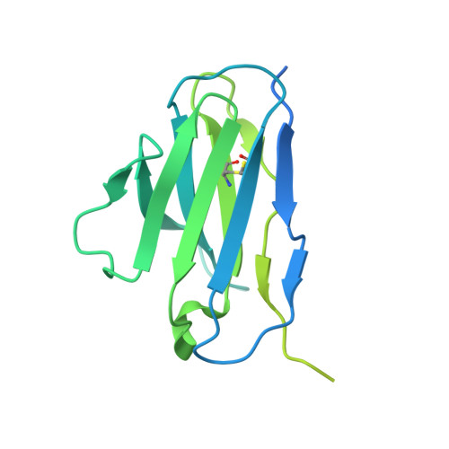
Cryo-EM structure of GluN1-2A NMDAR in complex with human Fab2G7 in one fab conformation


Experimental Data Snapshot

  • Method: ELECTRON MICROSCOPY
  • Resolution: 4.30 Å
  • Aggregation State: PARTICLE 
  • Reconstruction Method: SINGLE PARTICLE 

wwPDB Validation   3D Report Full Report


This is version 1.2 of the entry. See complete history


Literature

Structural basis for antibody-mediated NMDA receptor clustering and endocytosis in autoimmune encephalitis.

Wang, H.Xie, C.Deng, B.Ding, J.Li, N.Kou, Z.Jin, M.He, J.Wang, Q.Wen, H.Zhang, J.Zhou, Q.Chen, S.Chen, X.Yuan, T.F.Zhu, S.

(2024) Nat Struct Mol Biol 

  • DOI: https://doi.org/10.1038/s41594-024-01387-3
  • Primary Citation of Related Structures:  
    8JIZ, 8JJ0, 8JJ1, 8JJ2

  • PubMed Abstract: 

    Antibodies against N-methyl-D-aspartate receptors (NMDARs) are most frequently detected in persons with autoimmune encephalitis (AE) and used as diagnostic biomarkers. Elucidating the structural basis of monoclonal antibody (mAb) binding to NMDARs would facilitate the development of targeted therapy for AE. Here, we reconstructed nanodiscs containing green fluorescent protein-fused NMDARs to label and sort individual immune B cells from persons with AE and further cloned and identified mAbs against NMDARs. This allowed cryo-electron microscopy analysis of NMDAR-Fab complexes, revealing that autoantibodies bind to the R1 lobe of the N-terminal domain of the GluN1 subunit. Small-angle X-ray scattering studies demonstrated NMDAR-mAb stoichiometry of 2:1 or 1:2, structurally suitable for mAb-induced clustering and endocytosis of NMDARs. Importantly, these mAbs reduced the surface NMDARs and NMDAR-mediated currents, without tonically affecting NMDAR channel gating. These structural and functional findings imply that the design of neutralizing antibody binding to the R1 lobe of NMDARs represents a potential therapy for AE treatment.


  • Organizational Affiliation

    Institute of Neuroscience, CAS Center for Excellence in Brain Science and Intelligence Technology, Chinese Academy of Sciences, Shanghai, China.


Macromolecules
Find similar proteins by:  (by identity cutoff)  |  3D Structure
Entity ID: 1
MoleculeChains Sequence LengthOrganismDetailsImage
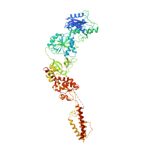
Glutamate receptor ionotropic, NMDA 2A
A, C
841Homo sapiensMutation(s): 0 
Gene Names: GRIN2ANMDAR2A
UniProt & NIH Common Fund Data Resources
Find proteins for Q12879 (Homo sapiens)
Go to UniProtKB:  Q12879
PHAROS:  Q12879
GTEx:  ENSG00000183454 
Glycosylation
Glycosylation Sites: 1Go to GlyGen: Q12879-1
Sequence Annotations
Expand
  • Reference Sequence
Find similar proteins by:  (by identity cutoff)  |  3D Structure
Entity ID: 2
MoleculeChains Sequence LengthOrganismDetailsImage
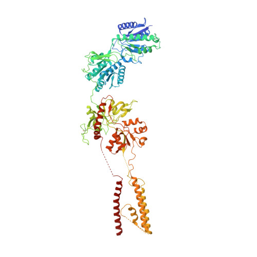
Glutamate receptor ionotropic, NMDA 1
B, D
847Homo sapiensMutation(s): 0 
Gene Names: GRIN1NMDAR1
UniProt & NIH Common Fund Data Resources
Find proteins for Q05586 (Homo sapiens)
Go to UniProtKB:  Q05586
PHAROS:  Q05586
GTEx:  ENSG00000176884 
Glycosylation
Glycosylation Sites: 4Go to GlyGen: Q05586-1
Sequence Annotations
Expand
  • Reference Sequence
Find similar proteins by:  (by identity cutoff)  |  3D Structure
Entity ID: 3
MoleculeChains Sequence LengthOrganismDetailsImage
Fab2G7 Heavy Chain253Homo sapiensMutation(s): 0 
Sequence Annotations
Expand
  • Reference Sequence
Find similar proteins by:  (by identity cutoff)  |  3D Structure
Entity ID: 4
MoleculeChains Sequence LengthOrganismDetailsImage
Fab2G7 Light Chain234Homo sapiensMutation(s): 0 
Sequence Annotations
Expand
  • Reference Sequence
Oligosaccharides

Help

Entity ID: 5
MoleculeChains Length2D Diagram Glycosylation3D Interactions
2-acetamido-2-deoxy-beta-D-glucopyranose-(1-4)-2-acetamido-2-deoxy-beta-D-glucopyranose
G
2N-Glycosylation
Glycosylation Resources
GlyTouCan:  G42666HT
GlyCosmos:  G42666HT
GlyGen:  G42666HT
Small Molecules
Ligands 1 Unique
IDChains Name / Formula / InChI Key2D Diagram3D Interactions
NAG (Subject of Investigation/LOI)
Query on NAG

Download Ideal Coordinates CCD File 
H [auth A]
I [auth B]
J [auth B]
K [auth B]
L [auth B]
H [auth A],
I [auth B],
J [auth B],
K [auth B],
L [auth B],
M [auth D],
N [auth D],
O [auth D]
2-acetamido-2-deoxy-beta-D-glucopyranose
C8 H15 N O6
OVRNDRQMDRJTHS-FMDGEEDCSA-N
Experimental Data & Validation

Experimental Data

  • Method: ELECTRON MICROSCOPY
  • Resolution: 4.30 Å
  • Aggregation State: PARTICLE 
  • Reconstruction Method: SINGLE PARTICLE 
EM Software:
TaskSoftware PackageVersion
RECONSTRUCTIONRELION

Structure Validation

View Full Validation Report


Entry History & Funding Information

Deposition Data


Funding OrganizationLocationGrant Number
Other governmentChina2022ZD0212700
Other governmentChinaLG 202106-02
Other governmentChina2017YFA0505700
Other governmentChinaXDBS01020000
Other governmentChina2018SHZDZX05

Revision History  (Full details and data files)

  • Version 1.0: 2024-06-05
    Type: Initial release
  • Version 1.1: 2024-09-11
    Changes: Data collection, Database references
  • Version 1.2: 2024-10-23
    Changes: Data collection, Structure summary