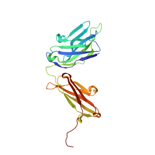
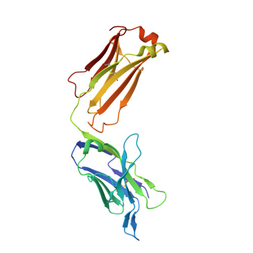
8JEQ

Crystal structure of Tiragolumab


Experimental Data Snapshot

  • Method: X-RAY DIFFRACTION
  • Resolution: 1.96 Å
  • R-Value Free: 0.222 
  • R-Value Work: 0.186 
  • R-Value Observed: 0.188 

Starting Model: experimental
View more details

wwPDB Validation   3D Report Full Report


This is version 1.3 of the entry. See complete history


Literature

Structural insights into the unique pH-responsive characteristics of the anti-TIGIT therapeutic antibody Ociperlimab.

Sun, J.Zhang, X.Xue, L.Cheng, L.Zhang, J.Chen, X.Shen, Z.Li, K.Wang, L.Huang, C.Song, J.

(2024) Structure 32: 550

  • DOI: https://doi.org/10.1016/j.str.2024.02.009
  • Primary Citation of Related Structures:  
    8JEL, 8JEN, 8JEO, 8JEP, 8JEQ

  • PubMed Abstract: 

    TIGIT is mainly expressed on T cells and is an inhibitory checkpoint receptor that binds to its ligand PVR in the tumor microenvironment. Anti-TIGIT monoclonal antibodies (mAbs) such as Ociperlimab and Tiragolumab block the TIGIT-PVR interaction and are in clinical development. However, the molecular blockade mechanism of these mAbs remains elusive. Here, we report the crystal structures of TIGIT in complex with Ociperlimab_Fab and Tiragolumab_Fab revealing that both mAbs bind TIGIT with a large steric clash with PVR. Furthermore, several critical epitopic residues are identified. Interestingly, the binding affinity of Ociperlimab toward TIGIT increases approximately 17-fold when lowering the pH from 7.4 to 6.0. Our structure shows a strong electrostatic interaction between ASP103 HCDR3 and HIS76 TIGIT explaining the pH-responsive mechanism of Ociperlimab. In contrast, Tiragolumab does not show an acidic pH-dependent binding enhancement. Our results provide valuable information that could help to improve the efficacy of therapeutic antibodies for cancer treatment.


  • Organizational Affiliation

    Department of Biologics, BeiGene (Beijing) Co., Ltd, Beijing, China.


Macromolecules
Find similar proteins by:  (by identity cutoff)  |  3D Structure
Entity ID: 1
MoleculeChains Sequence LengthOrganismDetailsImage
antibody heavy chain235Homo sapiensMutation(s): 0 
Entity Groups  
Sequence Clusters30% Identity50% Identity70% Identity90% Identity95% Identity100% Identity
Sequence Annotations
Expand
  • Reference Sequence
Find similar proteins by:  (by identity cutoff)  |  3D Structure
Entity ID: 2
MoleculeChains Sequence LengthOrganismDetailsImage
antibody light chain220Homo sapiensMutation(s): 0 
Entity Groups  
Sequence Clusters30% Identity50% Identity70% Identity90% Identity95% Identity100% Identity
Sequence Annotations
Expand
  • Reference Sequence
Experimental Data & Validation

Experimental Data

  • Method: X-RAY DIFFRACTION
  • Resolution: 1.96 Å
  • R-Value Free: 0.222 
  • R-Value Work: 0.186 
  • R-Value Observed: 0.188 
  • Space Group: P 21 21 21
Unit Cell:
Length ( Å )Angle ( ˚ )
a = 60.72α = 90
b = 89.05β = 90
c = 104.23γ = 90
Software Package:
Software NamePurpose
PHENIXrefinement
Aimlessdata scaling
xia2data reduction
PHASERphasing

Structure Validation

View Full Validation Report


Entry History & Funding Information

Deposition Data


Funding OrganizationLocationGrant Number
Not funded--

Revision History  (Full details and data files)

  • Version 1.0: 2024-02-28
    Type: Initial release
  • Version 1.1: 2024-03-20
    Changes: Database references
  • Version 1.2: 2024-05-22
    Changes: Database references
  • Version 1.3: 2024-11-13
    Changes: Structure summary