


Crystal structure of Procerain-B from Calotropis gigantea
Kumar, A., Jamdar, S.N., Srivastava, G., Makde, R.D.To be published.
Experimental Data Snapshot
Starting Model: experimental
View more details
wwPDB Validation 3D Report Full Report

Entity ID: 1 | |||||
|---|---|---|---|---|---|
| Molecule | Chains | Sequence Length | Organism | Details | Image |
| Procerain B | 339 | Calotropis | Mutation(s): 0 | 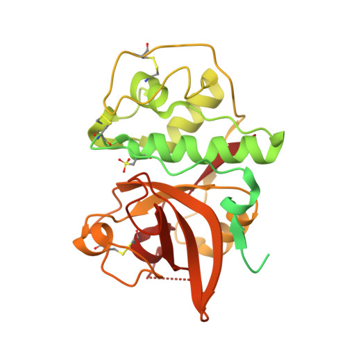 | |
UniProt | |||||
Find proteins for A0A0A0Q2K8 (Calotropis procera) Explore A0A0A0Q2K8 Go to UniProtKB: A0A0A0Q2K8 | |||||
Entity Groups | |||||
| Sequence Clusters | 30% Identity50% Identity70% Identity90% Identity95% Identity100% Identity | ||||
| UniProt Group | A0A0A0Q2K8 | ||||
Sequence AnnotationsExpand | |||||
| |||||
| Modified Residues 1 Unique | |||||
|---|---|---|---|---|---|
| ID | Chains | Type | Formula | 2D Diagram | Parent |
| OCS Query on OCS | A, B | L-PEPTIDE LINKING | C3 H7 N O5 S |  | CYS |
| Length ( Å ) | Angle ( ˚ ) |
|---|---|
| a = 31.426 | α = 90 |
| b = 91.466 | β = 90.019 |
| c = 64.392 | γ = 90 |
| Software Name | Purpose |
|---|---|
| PHENIX | refinement |
| XDS | data reduction |
| Aimless | data scaling |
| PHASER | phasing |
| PHENIX | model building |
| Coot | model building |
| Funding Organization | Location | Grant Number |
|---|---|---|
| Other government | India | -- |


