Crystal structure of the 3-ketodihydrosphingosine reductase TSC10 from Cryptococcus neoformans.
Zhao, P., Zhuang, Z., Guan, X., Yang, J., Wang, W., Kuang, Z.(2023) Biochem Biophys Res Commun 670: 73-78
- PubMed: 37285720 Search on PubMed
- DOI: https://doi.org/10.1016/j.bbrc.2023.05.109
- Primary Citation Related Structures:
8JAT - PubMed Abstract:
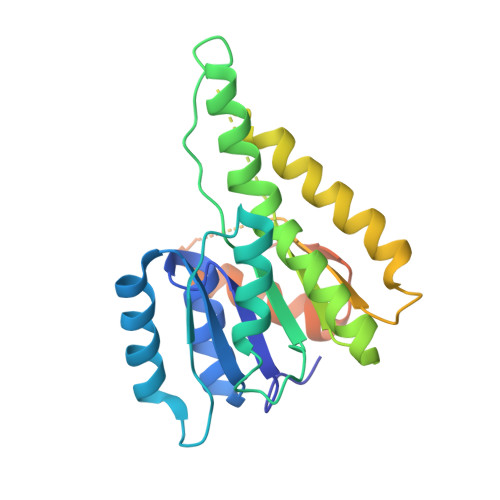
The second step in the de novo sphingolipid biosynthesis is the reduction of 3-ketodihydrosphingosine by 3-ketodihydrosphingosine reductase (KDSR) to produce dihydrosphingosine (sphinganine). Fungal TSC10 and mammalian KDSR (also named FVT-1) proteins are the enzymes responsible for this process and they belong to the short-chain dehydrogenase/reductase (SDR) superfamily. Albeit that both fungal and mammalian 3-ketodihydrosphingosine reductases were identified more than a decade ago, no structure of these enzymes from any species has been experimentally determined. Here we report the crystal structure of the catalytic domain of TSC10 from Cryptococcus neoformans in complex with NADPH. cnTSC10 adopts a Rossmann fold with a central seven-stranded β-sheet flanked by α-helices on both sides. Several regions are disordered that include the segment connecting the serine and tyrosine residues of the catalytic triad, the so-called 'substrate loop', and the C-terminal region that often participates in homo-tetramerization in other SDRs. In addition, the cofactor NADPH is not fully ordered. These structural features indicate that the catalytic site of cnTSC10 possesses significant flexibility. cnTSC10 is predominantly dimeric in solution while a minor portion of the protein forms homo-tetramer. The crystal structure reveals that the homo-dimer interface involves both hydrophobic and hydrophilic interactions mediated by helices α4 and α5, as well as the loop connecting strand β4 and helix α4. Because residues forming hydrogen bonds and salt bridges in the dimer interface are not conserved between fungal TSC10 and mammalian KDSR proteins, it might be possible to develop inhibitors that selectively target fungal TSC10 dimerization.
- Department of Cell Biology, College of Life Science and Technology, Jinan University, Guangzhou, 510632, China; Guangdong Provincial Key Laboratory of Bioengineering Medicine, Guangzhou, 510632, China; Guangdong Provincial Biotechnology Drug & Engineering Technology Research Center, Guangzhou, 510632, China; National Engineering Research Center of Genetic Medicine, Guangzhou, 510632, China.
Organizational Affiliation: