8J62 | pdb_00008j62

Cryo-EM structure of APOBEC3G-Vif complex


Experimental Data Snapshot

  • Method: ELECTRON MICROSCOPY
  • Resolution: 2.50 Å
  • Aggregation State: PARTICLE 
  • Reconstruction Method: SINGLE PARTICLE 

Starting Model: experimental
View more details

wwPDB Validation 3D Report Full Report

Validation slider image for 8J62

This is version 1.1 of the entry. See complete history

Literature

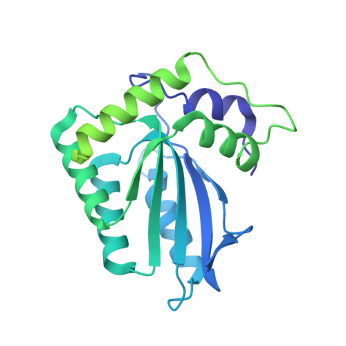
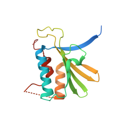
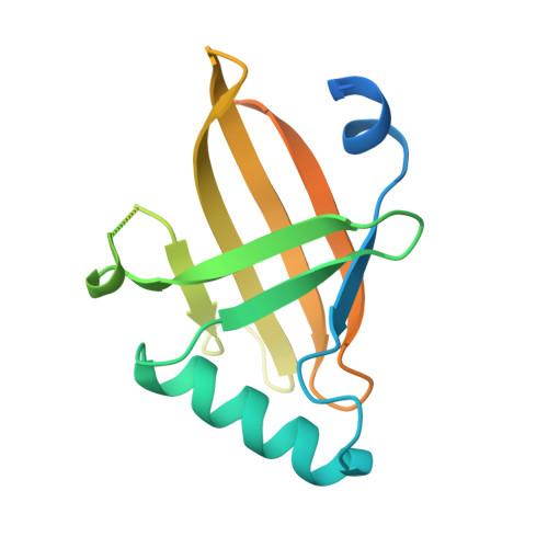
Structural insights into RNA bridging between HIV-1 Vif and antiviral factor APOBEC3G.

Kouno, T.Shibata, S.Shigematsu, M.Hyun, J.Kim, T.G.Matsuo, H.Wolf, M.

(2023) Nat Commun 14: 4037-4037

  • DOI: https://doi.org/10.1038/s41467-023-39796-5
  • Primary Citation Related Structures: 
    8H0I, 8J62

  • PubMed Abstract: 

    Great effort has been devoted to discovering the basis of A3G-Vif interaction, the key event of HIV's counteraction mechanism to evade antiviral innate immune response. Here we show reconstitution of the A3G-Vif complex and subsequent A3G ubiquitination in vitro and report the cryo-EM structure of the A3G-Vif complex at 2.8 Å resolution using solubility-enhanced variants of A3G and Vif. We present an atomic model of the A3G-Vif interface, which assembles via known amino acid determinants. This assembly is not achieved by protein-protein interaction alone, but also involves RNA. The cryo-EM structure and in vitro ubiquitination assays identify an adenine/guanine base preference for the interaction and a unique Vif-ribose contact. This establishes the biological significance of an RNA ligand. Further assessment of interactions between A3G, Vif, and RNA ligands show that the A3G-Vif assembly and subsequent ubiquitination can be controlled by amino acid mutations at the interface or by polynucleotide modification, suggesting that a specific chemical moiety would be a promising pharmacophore to inhibit the A3G-Vif interaction.


  • Organizational Affiliation
    • Molecular Cryo-Electron Microscopy Unit, Okinawa Institute of Science and Technology Graduate University, 1919-1 Tancha, Onna-son, Okinawa, 904-0495, Japan. 1st.soluble.fla3g@gmail.com.

Macromolecule Content 

  • Total Structure Weight: 247.42 kDa 
  • Atom Count: 10,448 
  • Modeled Residue Count: 1,220 
  • Deposited Residue Count: 2,006 
  • Unique protein chains: 3
  • Unique nucleic acid chains: 1

Macromolecules


Find similar proteins by:|  3D Structure
Entity ID: 1
MoleculeChains  Sequence LengthOrganismDetailsImage
APOBEC3GA,
G [auth B]
371Homo sapiensMutation(s): 0 
EC: 3.5.4.38
UniProt & NIH Common Fund Data Resources
Find proteins for Q9HC16 (Homo sapiens)
Explore Q9HC16 
Go to UniProtKB:  Q9HC16
PHAROS:  Q9HC16
GTEx:  ENSG00000239713 
Entity Groups
Sequence Clusters30% Identity50% Identity70% Identity90% Identity95% Identity100% Identity
UniProt GroupQ9HC16
Sequence Annotations
Expand
Reference Sequence
Find similar proteins by:|  3D Structure
Entity ID: 2
MoleculeChains  Sequence LengthOrganismDetailsImage
Viral infectivity factorB [auth C],
D [auth E],
H [auth G],
J [auth I]
150Human immunodeficiency virus 1Mutation(s): 0 
UniProt
Find proteins for P12504 (Human immunodeficiency virus type 1 group M subtype B (isolate NY5))
Explore P12504 
Go to UniProtKB:  P12504
Entity Groups
Sequence Clusters30% Identity50% Identity70% Identity90% Identity95% Identity100% Identity
UniProt GroupP12504
Sequence Annotations
Expand
Reference Sequence
Find similar proteins by:|  3D Structure
Entity ID: 3
MoleculeChains  Sequence LengthOrganismDetailsImage
Core binding factor betaC [auth D],
E [auth F],
I [auth H],
K [auth J]
156Homo sapiensMutation(s): 0 
UniProt & NIH Common Fund Data Resources
Find proteins for Q13951 (Homo sapiens)
Explore Q13951 
Go to UniProtKB:  Q13951
PHAROS:  Q13951
GTEx:  ENSG00000067955 
Entity Groups
Sequence Clusters30% Identity50% Identity70% Identity90% Identity95% Identity100% Identity
UniProt GroupQ13951
Sequence Annotations
Expand
Reference Sequence
Find similar nucleic acids by:  Sequence
Entity ID: 4
MoleculeChains LengthOrganismImage
RNA (5'-R(*CP*GP*GP*UP*UP*GP*AP*UP*UP*GP*UP*UP*UP*UP*AP*AP*CP*AP*A)-3')F [auth X],
L [auth Y]
20synthetic construct
Sequence Annotations
Expand
Reference Sequence

Experimental Data & Validation

Experimental Data

  • Method: ELECTRON MICROSCOPY
  • Resolution: 2.50 Å
  • Aggregation State: PARTICLE 
  • Reconstruction Method: SINGLE PARTICLE 
EM Software:
TaskSoftware PackageVersion
RECONSTRUCTIONRELION3.1.3
MODEL REFINEMENTPHENIX1.19.2

Structure Validation

View Full Validation Report


Entry History 

& Funding Information

Deposition Data


Funding OrganizationLocationGrant Number
Japan Agency for Medical Research and Development (AMED)JapanJP18am0101076

Revision History  (Full details and data files)

  • Version 1.0: 2023-07-19
    Type: Initial release
  • Version 1.1: 2024-07-03
    Changes: Data collection, Refinement description