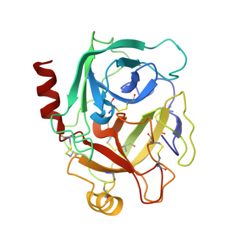
8IZH

Crystal structure of trypsin-aminoguanidine complex at 2.30 Angstroms resolution


Experimental Data Snapshot

  • Method: X-RAY DIFFRACTION
  • Resolution: 2.30 Å
  • R-Value Free: 0.295 
  • R-Value Work: 0.212 
  • R-Value Observed: 0.216 

Starting Model: experimental
View more details

wwPDB Validation   3D Report Full Report


Ligand Structure Quality Assessment 


This is version 1.1 of the entry. See complete history


Literature

Structural basis for the binding of famotidine, cimetidine, guanidine, and pimagedine with serine protease.

Ahmad, M.S.Kalam, N.Akbar, Z.Shah, N.Rasheed, S.Choudhary, M.I.

(2024) Biochem Biophys Res Commun 733: 150603-150603

  • DOI: https://doi.org/10.1016/j.bbrc.2024.150603
  • Primary Citation of Related Structures:  
    8IYV, 8IZH, 8IZI, 8IZK

  • PubMed Abstract: 

    Serine proteases are among the important groups of enzymes having significant roles in cell biology. Trypsin is a representative member of the serine superfamily of enzymes, produced by acinar cells of pancreas. It is a validated drug target for various ailments including pancreatitis and colorectal cancer. Premature activation of trypsin is involved in the lysis of pancreatic tissues, which causes pancreatitis. It is also reported to be involved in colorectal carcinoma by activating other proteases, such as matrix metalloproteinase (MMPs). The development of novel trypsin inhibitors with good pharmacokinetic properties could play important roles in pharmaceutical sciences. This study reports the crystal structures of bovine pancreatic trypsin with four molecules; cimetidine, famotidine, pimagedine, and guanidine. These compounds possess binding affinity towards the active site (S1) of trypsin. The structures of all four complexes provided insight of the binding of four different ligands, as well as the dynamics of the active site towards the bind with different size ligands. This study might be helpful in designing of new potent inhibitors of trypsin and trypsin like serine proteases.


  • Organizational Affiliation

    Dr. Panjwani Center for Molecular Medicine and Drug Research, International Center for Chemical and Biological Sciences, University of Karachi, Karachi, 75270, Pakistan; H.E.J. Research Institute of Chemistry, International Center for Chemical and Biological Sciences, University of Karachi, Karachi, 75270, Pakistan. Electronic address: shoaib.hej@gmail.com.


Macromolecules
Find similar proteins by:  (by identity cutoff)  |  3D Structure
Entity ID: 1
MoleculeChains Sequence LengthOrganismDetailsImage
Cationic trypsin223Bos taurusMutation(s): 0 
EC: 3.4.21.4
UniProt
Find proteins for P00760 (Bos taurus)
Explore P00760 
Go to UniProtKB:  P00760
Entity Groups  
Sequence Clusters30% Identity50% Identity70% Identity90% Identity95% Identity100% Identity
UniProt GroupP00760
Sequence Annotations
Expand
  • Reference Sequence
Small Molecules
Ligands 5 Unique
IDChains Name / Formula / InChI Key2D Diagram3D Interactions
SO4
Query on SO4

Download Ideal Coordinates CCD File 
E [auth A]SULFATE ION
O4 S
QAOWNCQODCNURD-UHFFFAOYSA-L
GOL
Query on GOL

Download Ideal Coordinates CCD File 
J [auth A],
K [auth A]
GLYCEROL
C3 H8 O3
PEDCQBHIVMGVHV-UHFFFAOYSA-N
AGU (Subject of Investigation/LOI)
Query on AGU

Download Ideal Coordinates CCD File 
F [auth A],
G [auth A],
H [auth A],
I [auth A]
AMINOGUANIDINE
C H6 N4
HAMNKKUPIHEESI-UHFFFAOYSA-N
CA
Query on CA

Download Ideal Coordinates CCD File 
B [auth A]CALCIUM ION
Ca
BHPQYMZQTOCNFJ-UHFFFAOYSA-N
CL
Query on CL

Download Ideal Coordinates CCD File 
C [auth A],
D [auth A]
CHLORIDE ION
Cl
VEXZGXHMUGYJMC-UHFFFAOYSA-M
Experimental Data & Validation

Experimental Data

  • Method: X-RAY DIFFRACTION
  • Resolution: 2.30 Å
  • R-Value Free: 0.295 
  • R-Value Work: 0.212 
  • R-Value Observed: 0.216 
  • Space Group: P 21 21 21
Unit Cell:
Length ( Å )Angle ( ˚ )
a = 54.59α = 90
b = 58.248β = 90
c = 66.326γ = 90
Software Package:
Software NamePurpose
REFMACrefinement
PROTEUM PLUSdata reduction
Aimlessdata scaling
MOLREPphasing

Structure Validation

View Full Validation Report


Ligand Structure Quality Assessment 


Entry History & Funding Information

Deposition Data


Funding OrganizationLocationGrant Number
Not fundedPakistan--

Revision History  (Full details and data files)

  • Version 1.0: 2024-09-18
    Type: Initial release
  • Version 1.1: 2024-11-13
    Changes: Structure summary