8IXQ | pdb_00008ixq

Structure of glycosyltransferase LmbT in complex with GDP and ergothioneine


Experimental Data Snapshot

  • Method: X-RAY DIFFRACTION
  • Resolution: 2.10 Å
  • R-Value Free: 
    0.221 (Depositor), 0.221 (DCC) 
  • R-Value Work: 
    0.184 (Depositor), 0.184 (DCC) 
  • R-Value Observed: 
    0.184 (Depositor) 

Starting Model: experimental
View more details

wwPDB Validation 3D Report Full Report

Validation slider image for 8IXQ

Ligand Structure Quality Assessment 


This is version 1.2 of the entry. See complete history

Literature

Structure-Function Analysis of the S-Glycosylation Reaction in the Biosynthesis of Lincosamide Antibiotics.

Mori, T.Sun, X.Kadlcik, S.Janata, J.Abe, I.

(2023) Angew Chem Int Ed Engl 62: e202304989-e202304989

  • DOI: https://doi.org/10.1002/anie.202304989
  • Primary Citation Related Structures: 
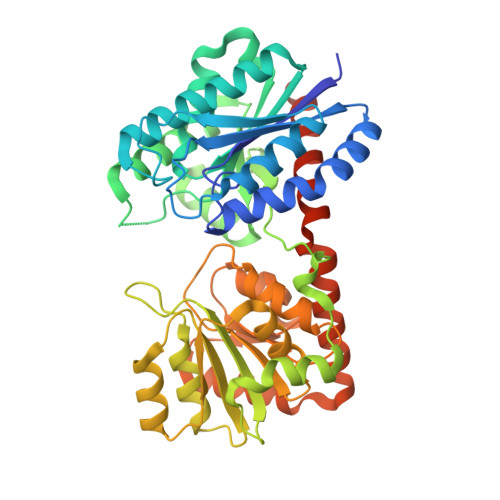
    8IXP, 8IXQ

  • PubMed Abstract: 

    The S-glycosyltransferase LmbT, involved in the biosynthesis of lincomycin A, is the only known enzyme that catalyzes the enzymatic incorporation of rare amino acid L-ergothioneine (EGT) into secondary metabolites. Here, we show the structure and function analyses of LmbT. Our in vitro analysis of LmbT revealed that the enzyme shows promiscuous substrate specificity toward nitrogenous base moieties in the generation of unnatural nucleotide diphosphate (NDP)-D-α-D-lincosamides. Furthermore, the X-ray crystal structures of LmbT in its apo form and in complex with substrates indicated that the large conformational changes of the active site occur upon binding of the substrates, and that EGT is strictly recognized by salt-bridge and cation-π interactions with Arg260 and Trp101, respectively. The structure of LmbT in complex with its substrates, the docking model with the EGT-S-conjugated lincosamide, and the structure-based site-directed mutagenesis analysis revealed the structural details of the LmbT-catalyzed S N 2-like S-glycosylation reaction with EGT.


  • Organizational Affiliation
    • Graduate School of Pharmaceutical Sciences, The University of Tokyo, 7-3-1 Hongo, Bunkyo-ku, Tokyo, 113-0033, Japan.

Macromolecule Content 

  • Total Structure Weight: 196.98 kDa 
  • Atom Count: 14,312 
  • Modeled Residue Count: 1,707 
  • Deposited Residue Count: 1,804 
  • Unique protein chains: 1

Macromolecules

Find similar proteins by:|  3D Structure
Entity ID: 1
MoleculeChains  Sequence LengthOrganismDetailsImage
Glycosyltransferase
A, B, C, D
451Streptomyces lincolnensisMutation(s): 0 
Gene Names: GJU35_01490
UniProt
Find proteins for A9Y8T1 (Streptomyces lincolnensis)
Explore A9Y8T1 
Go to UniProtKB:  A9Y8T1
Entity Groups
Sequence Clusters30% Identity50% Identity70% Identity90% Identity95% Identity100% Identity
UniProt GroupA9Y8T1
Sequence Annotations
Expand
Reference Sequence

Experimental Data & Validation

Experimental Data

  • Method: X-RAY DIFFRACTION
  • Resolution: 2.10 Å
  • R-Value Free:  0.221 (Depositor), 0.221 (DCC) 
  • R-Value Work:  0.184 (Depositor), 0.184 (DCC) 
  • R-Value Observed: 0.184 (Depositor) 
Space Group: P 21 21 21
Unit Cell:
Length ( Å )Angle ( ˚ )
a = 97.634α = 90
b = 139.484β = 90
c = 158.748γ = 90
Software Package:
Software NamePurpose
PHENIXrefinement
XDSdata reduction
Aimlessdata scaling
PHASERphasing

Structure Validation

View Full Validation Report


Ligand Structure Quality Assessment 


Entry History 

& Funding Information

Deposition Data


Funding OrganizationLocationGrant Number
Not funded--

Revision History  (Full details and data files)

  • Version 1.0: 2024-04-03
    Type: Initial release
  • Version 1.1: 2024-10-23
    Changes: Database references, Structure summary
  • Version 1.2: 2024-10-30
    Changes: Database references