


Crystal structure of peptidyl-tRNA hydrolase mutant from Enterococcus faecium
Pandey, R., Tripathi, S., Lanka, A.K., Zohib, M., Pal, R.K., Arora, A.To be published.
Experimental Data Snapshot
Starting Model: experimental
View more details
wwPDB Validation 3D Report Full Report

Entity ID: 1 | |||||
|---|---|---|---|---|---|
| Molecule | Chains | Sequence Length | Organism | Details | Image |
| Peptidyl-tRNA hydrolase | 186 | Enterococcus faecium | Mutation(s): 1 Gene Names: pth EC: 3.1.1.29 | 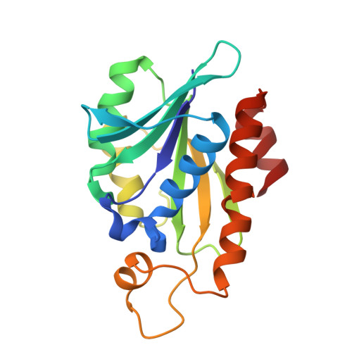 | |
UniProt | |||||
Entity Groups | |||||
| Sequence Clusters | 30% Identity50% Identity70% Identity90% Identity95% Identity100% Identity | ||||
| UniProt Group | A0A133CPV0 | ||||
Sequence AnnotationsExpand | |||||
Reference Sequence | |||||
| Ligands 1 Unique | |||||
|---|---|---|---|---|---|
| ID | Chains | Name / Formula / InChI Key | 2D Diagram | 3D Interactions | |
| GOL Download:Ideal Coordinates CCD File | C [auth A], D [auth B] | GLYCEROL C3 H8 O3 PEDCQBHIVMGVHV-UHFFFAOYSA-N |  | ||
| Length ( Å ) | Angle ( ˚ ) |
|---|---|
| a = 130.739 | α = 90 |
| b = 49.435 | β = 107.868 |
| c = 65.5 | γ = 90 |
| Software Name | Purpose |
|---|---|
| REFMAC | refinement |
| HKL-2000 | data scaling |
| HKL-2000 | data reduction |
| MOLREP | phasing |
| Funding Organization | Location | Grant Number |
|---|---|---|
| Council of Scientific & Industrial Research (CSIR) | India | CSIR FBR MLP 2029 |
| Department of Biotechnology (DBT, India) | India | BT/PR31893/MED/29/1390/2019 |


