Characterization of the extended substrate spectrum of the class A beta-lactamase CESS-1 from Stenotrophomonas sp. and structure-based investigation into its substrate preference.
Jeong, B.G., Kim, M.Y., Jeong, C.S., Do, H., Hwang, J., Lee, J.H., Cha, S.S.(2024) Int J Antimicrob Agents 63: 107171-107171
- PubMed: 38588869 Search on PubMed
- DOI: https://doi.org/10.1016/j.ijantimicag.2024.107171
- Primary Citation Related Structures:
8ISO, 8ISP, 8ISQ, 8ISR - PubMed Abstract:
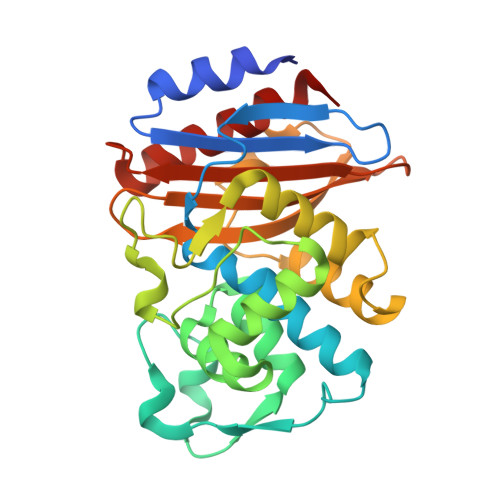
Stenotrophomonas spp. intrinsically resistant to many β-lactam antibiotics are found throughout the environment. CESS-1 identified in Stenotrophomonas sp. KCTC 12332 is an uncharacterized class A β-lactamase. The goal of this study was to reveal biochemical and structural characteristics of CESS-1. The hydrolytic activities of CESS-1 towards penicillins (penicillin G and ampicillin), cephalosporins (cephalexin, cefaclor, and cefotaxime), and carbapenems (imipenem and meropenem) was spectrophotometrically monitored. Structural information on E166Q mutants of CESS-1 acylated by cefaclor, cephalexin, or ampicillin were determined by X-ray crystallography. CESS-1 displayed hydrolytic activities toward penicillins and cephalosporins, with negligible activity toward carbapenems. Although cefaclor, cephalexin, and ampicillin have similar structures with identical R1 side chains, the catalytic parameters of CESS-1 toward them were distinct. The k cat values for cefaclor, cephalexin, and ampicillin were 1249.6 s -1 , 204.3 s -1 , and 69.8 s -1 , respectively, with the accompanying K M values of 287.6 μM, 236.7 μM, and 28.8 μM, respectively. CESS-1 was able to discriminate between cefaclor and cephalexin with a single structural difference at C3 position: -Cl (cefaclor) and -CH 3 (cephalexin). Structural comparisons among three E166Q mutants of CESS-1 acylated by cefaclor, cephalexin, or ampicillin, revealed that cooperative positional changes in the R1 side chain of substrates and their interaction with the β5-β6 loop affect the distance between Asn170 and the deacylating water at the acyl-enzyme intermediate state. This is directly associated with the differential hydrolytic activities of CESS-1 toward the three structurally similar β-lactam antibiotics.
- Department of Chemistry & Nanoscience, Ewha Womans University, Seoul, Republic of Korea.
Organizational Affiliation: