8IMH | pdb_00008imh

Solution structure of the N terminal domain of MazE9 antitoxin (nMazE9) from Mycobacterium tuberculosis


Experimental Data Snapshot

  • Method: SOLUTION NMR
  • Conformers Calculated: 200 
  • Conformers Submitted: 20 
  • Selection Criteria: structures with the lowest energy 

wwPDB Validation 3D Report Full Report

Validation slider image for 8IMH

This is version 1.2 of the entry. See complete history

Literature

Insights into the solution structure and transcriptional regulation of the MazE9 antitoxin in Mycobacterium tuberculosis.

Roy, T.B.Sarma, S.P.

(2025) Proteins 93: 176-196

  • DOI: https://doi.org/10.1002/prot.26589
  • Primary Citation Related Structures: 
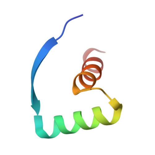
    8IMH

  • PubMed Abstract: 

    The present study endeavors to decode the details of the transcriptional autoregulation effected by the MazE9 antitoxin of the Mycobacterium tuberculosis MazEF9 toxin-antitoxin system. Regulation of this bicistronic operon at the level of transcription is a critical biochemical process that is key for the organism's stress adaptation and virulence. Here, we have reported the solution structure of the DNA binding domain of MazE9 and scrutinized the thermodynamic and kinetic parameters operational in its interaction with the promoter/operator region, specific to the mazEF9 operon. A HADDOCK model of MazE9 bound to its operator DNA has been calculated based on the information on interacting residues obtained from these studies. The thermodynamics and kinetics of the interaction of MazE9 with the functionally related mazEF6 operon indicate that the potential for intracellular cross-regulation is unlikely. An interesting feature of MazE9 is the cis ⇌ trans conformational isomerization of proline residues in the intrinsically disordered C-terminal domain of this antitoxin.


  • Organizational Affiliation
    • Molecular Biophysics Unit, Indian Institute of Science, Bangalore, Karnataka, India.

Macromolecule Content 

  • Total Structure Weight: 9.25 kDa 
  • Atom Count: 648 
  • Modeled Residue Count: 86 
  • Deposited Residue Count: 86 
  • Unique protein chains: 1

Macromolecules

Find similar proteins by:|  3D Structure
Entity ID: 1
MoleculeChains  Sequence LengthOrganismDetailsImage
Antitoxin MazE9
A, B
43Mycobacterium tuberculosis H37RvMutation(s): 0 
Gene Names: mazE9mazE-mt1Rv2801A
UniProt
Find proteins for P0CL61 (Mycobacterium tuberculosis (strain ATCC 25618 / H37Rv))
Explore P0CL61 
Go to UniProtKB:  P0CL61
Entity Groups
Sequence Clusters30% Identity50% Identity70% Identity90% Identity95% Identity100% Identity
UniProt GroupP0CL61
Sequence Annotations
Expand
Reference Sequence

Experimental Data & Validation

Experimental Data

  • Method: SOLUTION NMR
  • Conformers Calculated: 200 
  • Conformers Submitted: 20 
  • Selection Criteria: structures with the lowest energy 

Structure Validation

View Full Validation Report


Entry History 

& Funding Information

Deposition Data


Funding OrganizationLocationGrant Number
Department of Biotechnology (DBT, India)India--
Department of Science & Technology (DST, India)India--

Revision History  (Full details and data files)

  • Version 1.0: 2023-11-29
    Type: Initial release
  • Version 1.1: 2024-05-15
    Changes: Database references
  • Version 1.2: 2024-12-18
    Changes: Database references, Structure summary